Microfluidic Droplets: Miniature Bioreactors for Cells, Proteins, and Bacteria
Microfluidic droplets are femtolitre-to-nanolitre volumes of one fluid dispersed in a second, immiscible carrier fluid inside a microchannel [1]. Each droplet behaves as a sealed, independent compartment — in practice, a miniature bioreactor holding a single cell, enzyme or bacterium. This review covers what makes a droplet behave as a bioreactor, how biological material is encapsulated inside one, and which life-science applications the format unlocks. For the fluid physics of droplet formation itself, see our dedicated microfluidic droplet production method guide.
Last updated: August 2026.
What Are Microfluidic Droplets?
Microfluidic droplets are uniform volumes of a dispersed phase suspended in an immiscible continuous phase, formed inside microchannels typically tens to hundreds of micrometres wide. Individual droplet volumes span the femtolitre to nanolitre range, and each one is physically separated from its neighbours by the carrier fluid [1].
Because channel geometry and the two flow rates set the volume, a single microdevice produces millions of near-identical compartments per hour [2,3] — the reproducibility that separates droplet microfluidics from bulk emulsification. Figure 1 shows a classical microfluidic setup and a droplet-generation image from the EZ Drop chip [4].
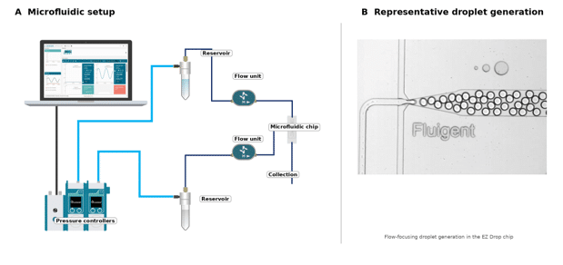
Figure 1. EZ Drop droplet-generation workflow. (A) Pressure-based microfluidic setup with the main components labeled directly beside each item: pressure controllers, reservoirs, flow units, microfluidic chip and collection. (B) Droplet formation in the EZ Drop flow-focusing chip. Source: adapted from Fluigent Droplet Generation Pack / EZ Drop [4].
Monodisperse Droplets and Why Uniformity Matters
Monodisperse droplets are populations whose diameters vary by only a few percent. Uniformity matters because droplet volume sets reagent concentration, incubation conditions and signal intensity, so any spread in size propagates directly into assay variance across the emulsion.
Droplet size and frequency depend on the stability of both flow rates: the operating diagram of drop size against flow-rate ratio is the basic design tool for any generator [2,5]. Pressure-driven flow control settles faster and shows lower flow-rate variation than the syringe-pump system tested in a dedicated droplet-generation comparison, which is why droplet size is a practical readout of fluidic stability [6,7].
Dispersed Phase and Continuous Phase
The dispersed phase forms the droplets: usually an aqueous buffer carrying cells, reagents or a hydrogel precursor. The continuous phase is the immiscible carrier — most often a fluorinated oil with a biocompatible surfactant [8]. Inverting the two phases produces oil-in-water droplets instead of water-in-oil. In the microfluidic setup shown in Figure 1A, the dispersed and continuous phases are delivered independently to the flow-focusing chip [4]. A published example distinguishing dispersed and continuous streams at the droplet-generation junction is shown in Figure 2 [9].
Phase selection governs biocompatibility, gas permeability and optical readout. Perfluorocarbon oils are widely used for cell work because gas-permeable carrier and storage systems allow encapsulated cells to survive and proliferate on chip [10].

Figure 2. Example of dispersed and continuous phases in a microfluidic droplet-generation device. Cells/collagen and alginate are introduced as dispersed-phase streams (solid arrows), while oil is introduced as the continuous phase (dotted arrows). From Liu et al. (2023), Frontiers in Bioengineering and Biotechnology, CC BY 4.0 [9].
Droplet Microfluidics vs Continuous-Flow Microfluidics
Continuous-flow microfluidics moves a single fluid phase through channels, so molecules diffuse and mix along the whole path. Droplet microfluidics compartmentalises the same chemistry into discrete volumes, eliminating cross-contamination and dispersion between samples [1,3].
The consequence is scale: a droplet emulsion holds millions of parallel conditions in a single tube. Both approaches are covered in our droplet and particle generation expertise hub.
Microfluidic Droplets as Miniature Bioreactors
A microfluidic droplet acts as a bioreactor when it confines one biological reaction — a growing cell, an expressed protein, a dividing bacterium — inside a defined, controlled and isolated volume. The droplet supplies the vessel, the medium and the boundary, at a scale six to nine orders of magnitude below a conventional culture flask.
The literature treats these compartments as controlled microbioreactors for single cells and small microbial populations rather than as passive containers, and complete cultivation and assay protocols are now run inside them [11].
Droplet microfluidics can also template cell-laden hydrogel microparticles, converting transient droplets into crosslinked, structured microenvironments whose material, architecture and fabrication can be tuned for cell culture, scale-up and biomedical applications [12] (Figure 3).
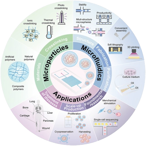
Figure 3. Overview of droplet microfluidics-powered hydrogel microparticles, including materials and crosslinking, microfluidic structure/fabrication, and biomedical applications. Source: Zheng et al. [12], Small (2024). © 2024 Wiley-VCH GmbH.
One Droplet, One Isolated Reaction Compartment
Isolation is the defining property. Surfactant-stabilised droplets do not exchange contents, so a secreted enzyme, a metabolite or a lysed cell’s RNA stays with its parent compartment throughout incubation, sorting and analysis [8,13].
That containment is what makes genotype-to-phenotype linkage possible. A cell and the product it makes remain physically paired, so a fluorescent readout can be traced back to the exact variant that produced it [13,14].
Controlled Microenvironments and Reagent Economy
Each droplet is a defined microenvironment: known volume, known starting concentration, uniform temperature. Reagent use falls in proportion to the volume — droplet platforms have been reported to run assays in over a thousandfold smaller volumes than microtitre plates [10].
The economics are documented. Agresti and colleagues screened roughly 10⁸ individual enzyme reactions in about ten hours using under 150 µL of total reagent — a thousandfold gain in speed and a millionfold reduction in cost against contemporary robotic screening [14].
Surfactants and the Droplet Interface
Surfactants adsorb at the oil-water interface and prevent coalescence when droplets are packed, stored or reinjected [8]. In biological workflows they must also stabilize the interface without significantly perturbing cells, enzymes, nucleic acids or fluorescence readouts.
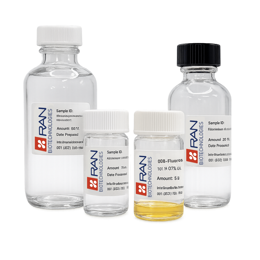
A current example is 008-FluoroSurfactant, developed by RAN Biotechnologies and available through Fluigent. It is designed for reproducible droplet formation, long-term emulsion stability, resistance to thermal and mechanical stress, and compatibility with enzymes, nucleic acids, fluorophores and cells [15]. Figure 4 illustrates stable droplets before and after PCR thermal cycling and extended incubation using 008-FluoroSurfactant [15]. Device surface chemistry remains equally important: wetting by the dispersed phase can destabilize droplet formation [16].

Figure 4. Droplet stability with 008-FluoroSurfactant developed by RAN Biotechnologies before and after PCR thermal cycling and incubation. Source: RAN Biotechnologies.
How Long Can Cells Survive Inside a Droplet?
Mammalian cells and microorganisms remain viable inside droplets for hours to days, depending on droplet volume, nutrient load, oxygen supply through the carrier oil, and the accumulation of metabolic waste in a closed compartment.
Human cell lines encapsulated in 660 pL droplets have been kept viable, recovered and recultivated after days of incubation, and even a multicellular organism has been maintained in the format [10]. Longer runs need larger droplets or periodic reagent addition (Figure 5).

Figure 5. HEK293T cell viability and spheroid formation in alginate microbeads at days 2, 6 and 10. Source: Rembotte et al. [17]; the image is also presented in the related spheroid-encapsulation case study.
How Microfluidic Droplets Are Generated
Droplets form where two immiscible streams meet in a microchannel and interfacial forces overcome viscous shear, pinching the dispersed phase into discrete volumes. Flow rates, channel geometry, viscosity and surfactant concentration together set droplet size and production frequency [3,18].
Generation Geometries: Flow-Focusing, Co-Flow, T-Junction
Three geometries dominate. T-junctions shear the dispersed phase at a perpendicular intersection [5]; flow-focusing forces it through a constriction between two continuous-phase streams [2]; co-flow generates droplets from a coaxial nozzle [16]. Each offers a different size-versus-frequency operating window.
Single vs Double Emulsions
Single emulsions contain one interface, typically water-in-oil (W/O) or oil-in-water (O/W). Double emulsions add a second interface – for example water-in-oil-in-water (W/O/W) – producing a core-shell compartment whose aqueous outer phase is compatible with standard flow cytometry and FACS sorting [15]. Figure 6 compares single-emulsion and double-emulsion generation in a nozzle-collection-capillary geometry and the resulting single-interface and core-shell droplet structures [19,20].
Full parameter maps, scaling laws and troubleshooting are covered in the microfluidic droplet production method article and in our droplet generation applications.

Figure 6. Representative single- and double-emulsion droplet formation in a nozzle-collection-capillary geometry(RayDrop developed by Secoya Technologies) . Left: single-interface droplets. Right: a core-shell double-emulsion droplet. Source: adapted from Fluigent application and product resources [4,27,36].
Encapsulation Strategies in Microfluidic Droplets
Encapsulation loads biological material into droplets at the moment of formation, by mixing it into the dispersed phase upstream of the junction. Payload dictates strategy: single cells, purified proteins, motile bacteria and gelling polymers each impose distinct constraints on droplet size, chemistry and downstream handling [21]. (Figure 7)

Figure 7. Representative microfluidic encapsulation examples: (A) PBMC cell encapsulation; (B) yeast encapsulation; (C) bacterial encapsulation; and (D) PLGA microbeads encapsulating lysozyme. Credits: (A) Fluigent/Secoya cell-encapsulation application note [22]; (B,C) Fluigent/Secoya/TU Delft [23], images courtesy of Marijke Luttik, Sagarika B. Govindaraju and Rinke van Tatenhove-Pel, TU Delft; (D) image by Secoya Technologies, source: Fluigent [24].
Cell and Single-Cell Encapsulation
Single-cell encapsulation isolates one cell per droplet so that its behaviour, secretions or transcriptome can be measured without population averaging [10,25]. Cell suspension density is tuned upstream to control how many cells enter each compartment. (Figure 7A)
Double emulsions are frequently preferred for cell work because the water-in-oil-in-water format is FACS-compatible [16]. See the application note on cell encapsulation in small double emulsions, or the dedicated cell encapsulation platform.
Poisson Loading and Encapsulation Efficiency
Cells arriving at a junction are randomly distributed, so occupancy follows Poisson statistics: pushing for a high proportion of singly occupied droplets also increases doublets [26]. Most workflows therefore dilute the suspension, accepting many empty droplets to keep multi-cell compartments rare.
Cell ordering can overcome the occupancy statistics associated with random Poisson loading. Hydrodynamic ordering in a high-aspect-ratio channel can synchronize cell arrival with droplet formation, increasing the fraction of single-cell droplets while reducing empty and multicell droplets [27]. Dean-flow-based inertial ordering in curved microchannels can similarly increase single-cell encapsulation efficiency above that expected from random loading [28].
Protein and Enzyme Encapsulation
Proteins and enzymes are encapsulated to run millions of parallel biochemical reactions, each in its own compartment [14]. Because the droplet volume is fixed and known, fluorescent product accumulation converts directly into a kinetic measurement per droplet. (Figure 7D)
The main technical risk is the interface: without an appropriate surfactant chemistry the oil–water boundary is not inert, and gene expression or enzyme assays inside the droplet are compromised [8].
Bacterial and Microbial Encapsulation
Encapsulating bacteria or yeast gives each clone a private culture vessel, allowing growth, antibiotic response or secreted activity to be scored strain by strain. This is the basis of droplet approaches to cultivating organisms that resist conventional plating [11]. (Figure 7B,C)
Droplet microfluidics is now applied across pathogen identification, antibiotic susceptibility testing, microbial physiology and strain improvement [11], and to resolving the composition and activity of complex microbiomes [29]. Method detail: microencapsulation of bacteria and yeast.
Hydrogels and Microcapsules
A gelling precursor — alginate, agarose, PEG or PLGA — can be encapsulated and crosslinked, converting the liquid droplet into a solid microsphere or core-shell microcapsule. Double emulsions are the standard template, with shell composition set by the middle phase [16].
These formats support cell therapy carriers, controlled release and 3D culture scaffolds. Our microcapsule generation white paper covers the chemistry and the fluidic setup. (Figure 8)

Figure 8. PLGA double emulsions and PLGA microcapsules after shell precipitation. Source: Fluigent, PLGA Microcapsule Synthesis [30]; the RayDrop Double Emulsion used in this workflow is developed and manufactured by Secoya Technologies.
| Payload | Typical droplet format | Illustrative size scale | Downstream readout |
|---|---|---|---|
| Mammalian / single cells | W/O or W/O/W | <90 µm for FACS-compatible DE workflows [31] | Imaging, secretion assay, FACS |
| Bacteria / yeast | W/O or W/O/W | ~42 µm in the cited DE example [23] | Growth, phenotype, FACS |
| Proteins / enzymes | W/O | Assay-dependent; typically pL–nL compartments [1,14] | Fluorescence / kinetic assay |
| Hydrogels / polymers | W/O, O/W or W/O/W | 230–312 µm in the cited PLGA microcapsule example [30] | 3D culture, delivery, recovery |
Applications of Droplet Bioreactors in the Life Sciences
Droplet bioreactors are used wherever an experiment requires many isolated biological reactions rather than one large one. The recurring pattern is the same across fields: compartmentalise, incubate, read out, and sort the rare positives [21].
High-Throughput Screening
Droplet screening evaluates libraries at rates unreachable by plate-based robotics, because each droplet replaces a well [25]. Fluorescence-activated droplet sorting then recovers hits from populations of 10⁷ to 10⁸ compartments, at rates up to about 2,000 droplets per second [13].
Single-Cell Analysis and Sequencing
Droplet barcoding pairs each cell with a uniquely tagged bead, allowing thousands of transcriptomes to be sequenced together while preserving cell of origin. Drop-seq demonstrated this on roughly 45,000 mouse retinal cells, resolving 39 cell types [32]; inDrop reported a parallel approach in the same issue [33].
Fluigent supplies a Drop-seq chip for this workflow [34]. (Figure 9)

Figure 9. Principle of the Drop-seq workflow: cell and barcoded microparticle co-encapsulation, cell lysis, RNA hybridization, reverse transcription, PCR, sequencing and analysis. Source: Fluigent, Droplet Sequencing: Drop-Seq Method [34]. The Drop-Seq flow-focusing chip described on the page is designed and developed by FlowJEM.
Directed Enzyme Evolution and Drug Discovery
Directed evolution needs genotype and phenotype kept together, which is exactly what a droplet enforces. Variant libraries are expressed, assayed and sorted in-droplet, then the winning genes are recovered and re-mutagenised for the next round [14,21].
Microbial Cultivation and Bioprocessing
Droplets let individual environmental isolates grow clonally in parallel, surfacing slow-growing or rare organisms that batch culture overgrows [11,29]. The same principle supports strain selection for production hosts before scale-up. (Figure 10)

Figure 10. E. coli culture in microfluidic droplets after 4 h and 20 h incubation, shown in bright-field and dark-field imaging. Source: Fluigent [35]. The study was conducted in collaboration with Dr Oksana Shvydkiv and her lab at the Leibniz Institute for Natural Product Research and Infection Biology.
Digital PCR and Diagnostics
Digital PCR partitions a sample across thousands of droplets so that target molecules are counted as positive or negative compartments, giving absolute quantification without a standard curve; commercial droplet systems process on the order of two million partitioned reactions per run [36]. Fluigent’s fluid handling for digital PCR covers OEM integration.
Drug Delivery and Microparticle Formulation
Monodisperse microspheres and microcapsules produced by droplet templating give tightly controlled release kinetics, because payload release depends on shell thickness and particle size — both set at the generation step [16,37].
Advantages Over Conventional Bioreactors
Compared with flasks, plates and stirred-tank vessels, droplet bioreactors trade absolute volume for parallelism and control. The advantage is not that a droplet does more, but that millions of droplets each do a little, independently and identically.
| Criterion | Conventional bioreactor / microplate | Microfluidic droplet bioreactor |
|---|---|---|
| Working volume | mL to L | fL to nL [1] |
| Parallel conditions | 96–1,536 wells per HTS plate [38] | 10⁶–10⁸ [14] |
| Reagent per test | µL–mL | pL–nL [10,14] |
| Cross-contamination | Handling-dependent | Prevented by the carrier phase [8] |
| Single-cell resolution | Rare, laborious | Native to the format [26,32] |
| Sorting of positives | Plate replication | Droplet sorting, ~2 kHz [13] |
Throughput, Volume and Cost
Reagent spend scales with compartment size, so shrinking the vessel by six orders of magnitude shrinks the consumables bill with it. The reported millionfold cost reduction in droplet enzyme screening comes from precisely this scaling [14], alongside roughly 500-fold higher throughput than microtitre plates in cell-based assays [10].
Limitations to Keep in Mind
Droplets are not universal. Long cultivations exhaust nutrients in a closed compartment, mid-experiment additions need dedicated hardware, and recovering material means breaking the emulsion. High-viscosity or particle-laden systems also generate less reliably [3,11].
Conclusion: Our Solutions for Droplet Bioreactors
Reliable droplet bioreactors need two things: a generation device that produces the target droplet format, and flow control stable enough to keep every droplet identical for the duration of the run. Fluigent supplies both, plus the assembled packages for common encapsulation workflows.
RayDrop: Single- and Double-Emulsion Generation (developed by Secoya Technologies)
The RayDrop is a nozzle-based droplet generator in which the dispersed phase never touches the device wall, removing the wetting problems that affect planar chips. It exists in a single-emulsion version and a double-emulsion version for core-shell and FACS-compatible work. [19]
Flow EZ and Flow Unit: Pressure-Driven Stability
Monodispersity depends on pulseless, fast-settling flow, since droplet size tracks the flow-rate ratio of the two phases [2]. The Flow EZ pressure controller drives both phases without the mechanical pulsation of a syringe drive, and the Flow Unit flow sensor closes the loop to hold droplet frequency constant across long runs [6].
Turnkey Encapsulation Platforms
Two assembled packages cover the most common needs: the cell encapsulation platform for FACS-compatible single-cell work, and the complex emulsion production platform for double emulsions and multi-layer microcapsules [31]. In the FACS application note, the platform was used to encapsulate fluorescent E. coli in W/O/W double emulsions before fluorescence-activated droplet sorting [39] (Figure 11).

Figure 11. FACS-compatible bacterial encapsulation workflow. (A) Microfluidic circuit of the standard FACS-compatible encapsulation platform, integrating pressure control for the continuous, shell and core phases with chip connection, collection and waste paths. (B) Microscopic observation of W/O/W double emulsions containing fluorescent E. coli, shown as a brightfield + GFP image stack (scale bar = 20 µm). Sources: Fluigent Encapsulation Platform for FACS [31] and Fluigent, A quick and efficient double encapsulation method for FACS-based droplet sorting, Figure 4A [39]. The FACS application was developed in partnership with TWB/TBI and ICEO-PICT collaborators.
Interested in integrating droplet microfluidics into your biological workflow?
Contact us to discuss your protocol.
Frequently Asked Questions
What is a microfluidic droplet bioreactor?
A microfluidic droplet bioreactor is a femtolitre-to-nanolitre droplet used as an isolated vessel for a single biological reaction. It supplies a defined volume, a controlled medium and a surfactant-stabilised boundary, so one cell, enzyme or microbe can be cultured and measured independently of all others [10,11].
How are cells encapsulated in microfluidic droplets?
Cells are suspended in the aqueous dispersed phase and carried to a droplet generation junction, where the immiscible carrier fluid pinches the stream into droplets. Suspension density is diluted so that occupancy follows Poisson statistics and most occupied droplets contain exactly one cell [26].
What is the difference between single and double emulsion droplets?
A single emulsion is a water-in-oil droplet with one interface. A double emulsion is water-in-oil-in-water: an aqueous core, an oil shell and an aqueous outer phase. The aqueous exterior makes double emulsions compatible with flow cytometry and standard FACS instruments [16].
Why are monodisperse droplets important for biological assays?
Droplet volume determines reagent concentration, incubation conditions and fluorescence signal. If droplet sizes vary, that variation appears directly in the assay readout and cannot be separated from real biological differences, degrading the statistics of any screen [1,2].
What equipment is needed to generate microfluidic droplets?
Three elements: a droplet generation device such as a RayDrop or a microfluidic chip, a flow control system for both phases, a camera visualization system and a surfactant-stabilised fluid pair [6,8,19].
Related Expertises
-
Microfluidics Case Studies Microfluidic Spheroid Encapsulation in Alginate Microbeads Using a Sacrificial Oil-Shell Method Read more
-
Microfluidics Case Studies Microfluidic Cell Encapsulation for Directed Evolution of Cellulose-Producing Microorganisms Read more
-
Expert Reviews: Basics of Microfluidics 10 Tips for Reliable Droplet Generation Read more
-
Microfluidics Case Studies Microfluidic Control of Complex Emulsions for Chemical Sensing Read more
-
Microfluidic Application Notes A quick and efficient double encapsulation method for FACS-based droplet sorting Read more
-
Microfluidics White Papers Double emulsion for the generation of microcapsules – a Review Read more
-
Microfluidic Application Notes What is the best method for Microencapsulation of Bacteria and Yeast in Small Double Emulsions? Read more
-
Microfluidic Application Notes Encapsulation of Cells In Small Double Emulsions Read more
-
Microfluidics White Papers Droplet-based Microfluidics – A Complete Guide Read more
-
Expert Reviews: Basics of Microfluidics High Throughput Single Cell Analysis Read more
-
Expert Reviews: Basics of Microfluidics Microfluidic Droplet Production Method Read more
References
[1] Teh S-Y, Lin R, Hung L-H, Lee AP. Droplet microfluidics. Lab on a Chip. 2008;8:198–220. doi:10.1039/b715524g
[2] Anna SL, Bontoux N, Stone HA. Formation of dispersions using “flow focusing” in microchannels. Applied Physics Letters. 2003;82(3):364–366. doi:10.1063/1.1537519
[3] Nan L, Zhang H, Weitz DA, Shum HC. Development and future of droplet microfluidics. Lab on a Chip. 2024;24(5):1135–1153. doi:10.1039/d3lc00729d
[4] Fluigent. Microfluidic Droplet Pack and EZ Drop easy droplet generation chip. Official product and application resources describing the EZ Drop flow-focusing chip, pressure-driven liquid handling, flow sensing, and monodisperse droplet generation. Fluigent website. Accessed 13 August 2026.
[5] Garstecki P, Fuerstman MJ, Stone HA, Whitesides GM. Formation of droplets and bubbles in a microfluidic T-junction — scaling and mechanism of break-up. Lab on a Chip. 2006;6(3):437–446.
[6] Fluigent. Flow EZ and FLOW UNIT. Official product documentation for pressure-based control, flow-rate measurement and DFC flow-rate regulation. Fluigent website. Accessed 12 August 2026.
[7] Fluigent. Microfluidic Droplet Generation Using Syringe Pumps and Pressure-Based Flow Controllers. Application note. Comparative measurements of flow-rate stability, droplet-size stability and response time using syringe pumps and Flow EZ pressure-based control. Fluigent website. Accessed 12 August 2026.
[8] Holtze C, Rowat AC, Agresti JJ, et al. Biocompatible surfactants for water-in-fluorocarbon emulsions. Lab on a Chip. 2008;8(10):1632–1639. doi:10.1039/b806706f
[9] Liu D, Xuanyuan T, Liu X, Fu W, Liu W. Massive and efficient encapsulation of single cells in monodisperse droplets and collagen–alginate microgels using a microfluidic device. Frontiers in Bioengineering and Biotechnology. 2023;11:1281375. doi:10.3389/fbioe.2023.1281375. CC BY 4.0.
[10] Clausell-Tormos J, Lieber D, Baret J-C, et al. Droplet-based microfluidic platforms for the encapsulation and screening of mammalian cells and multicellular organisms. Chemistry & Biology. 2008;15:427–437. doi:10.1016/j.chembiol.2008.04.004
[11] Kaminski TS, Scheler O, Garstecki P. Droplet microfluidics for microbiology: techniques, applications and challenges. Lab on a Chip. 2016;16(12):2168–2187. doi:10.1039/c6lc00367b
[12] Zheng F, Tian R, Lu H, Liang X, Shafiq M, Uchida S, Chen H, Ma M. Droplet Microfluidics Powered Hydrogel Microparticles for Stem Cell-Mediated Biomedical Applications. Small. 2024;20(42):e2401400. doi:10.1002/smll.202401400
[13] Baret J-C, Miller OJ, Taly V, et al. Fluorescence-activated droplet sorting (FADS): efficient microfluidic cell sorting based on enzymatic activity. Lab on a Chip. 2009;9(13):1850–1858. doi:10.1039/b902504a
[14] Agresti JJ, Antipov E, Abate AR, et al. Ultrahigh-throughput screening in drop-based microfluidics for directed evolution. PNAS. 2010;107(9):4004–4009. doi:10.1073/pnas.0910781107
[15] Fluigent / RAN Biotechnologies. 008-FluoroSurfactant – High-Performance Surfactant for Droplet Microfluidics. 008-FluoroSurfactant developed by RAN Biotechnologies. Fluigent website. Accessed 12 August 2026.
[16] Utada AS, Lorenceau E, Link DR, Kaplan PD, Stone HA, Weitz DA. Monodisperse double emulsions generated from a microcapillary device. Science. 2005;308(5721):537–541. doi:10.1126/science.1109164
[17] Rembotte L, Cappello J, Dewandre A, Mettler M, Septavaux J, Nassoy P, Scheid B. Sacrificial oil shell method for the generation of alginate microbeads adapted to multicellular spheroid culture. Lab on a Chip. 2026;26:711–724. doi:10.1039/D5LC00913H
[18] Fluigent. Microfluidic Droplet Production Method. Expertise review. Fluigent website. Accessed 12 August 2026.
[19] Fluigent. RayDrop Double Emulsion. Developed and manufactured by Secoya Technologies. Fluigent website. Accessed 12 August 2026.
[20] Fluigent. RayDrop Single Emulsion. Microfluidic single-emulsion droplet generator developed and manufactured by Secoya Technologies. Fluigent website. Accessed 12 August 2026.
[21] Shembekar N, Chaipan C, Utharala R, Merten CA. Droplet-based microfluidics in drug discovery, transcriptomics and high-throughput molecular genetics. Lab on a Chip. 2016;16:1314–1331. doi:10.1039/c6lc00249h
[22] Fluigent. Cell Encapsulation in Small Double Emulsions. Application note. Cell Encapsulation Platform developed and manufactured by Secoya Technologies. Fluigent website. Accessed 12 August 2026.
[23] Fluigent, Secoya Technologies, and TU Delft. Microencapsulation of Bacteria and Yeast in Small Double Emulsions. Application note. Images courtesy of Marijke Luttik, Sagarika B. Govindaraju and Rinke van Tatenhove-Pel, TU Delft. Fluigent website. Accessed 12 August 2026.
[24] Fluigent. Microfluidics in Drug Delivery: Techniques, Applications & Benefits. PLGA microbeads encapsulating lysozyme produced by Secoya Technologies. Fluigent website. Accessed 12 August 2026.
[25] Brouzes E, Medkova M, Savenelli N, et al. Droplet microfluidic technology for single-cell high-throughput screening. PNAS. 2009;106:14195–14200. doi:10.1073/pnas.0903542106
[26] Collins DJ, Neild A, deMello A, Liu AQ, Ai Y. The Poisson distribution and beyond: methods for microfluidic droplet production and single cell encapsulation. Lab on a Chip. 2015;15:3439–3459. doi:10.1039/c5lc00614g
[27] Edd JF, Di Carlo D, Humphry KJ, Köster S, Irimia D, Weitz DA, Toner M. Controlled encapsulation of single-cells into monodisperse picolitre drops. Lab on a Chip. 2008;8(8):1262–1264. doi:10.1039/b805456h
[28] Kemna EWM, Schoeman RM, Wolbers F, Vermes I, Weitz DA, van den Berg A. High-yield cell ordering and deterministic cell-in-droplet encapsulation using Dean flow in a curved microchannel. Lab on a Chip. 2012;12(16):2881–2887. doi:10.1039/c2lc00013j
[29] Xu Y, Wang Z, Li C, Tian S, Du W. Droplet microfluidics: unveiling the hidden complexity of the human microbiome. Lab on a Chip. 2025;25:1128. doi:10.1039/d4lc00877d
[30] Fluigent. Stable Monodispersed PLGA Microcapsules Synthesis. Application note. RayDrop Double Emulsion developed and manufactured by Secoya Technologies. Fluigent website. Accessed 12 August 2026.
[31] Fluigent. Encapsulation Platform for FACS; Microfluidic Complex Emulsion Production Platform. The FACS platform is developed and manufactured by Secoya using Fluigent flow-control equipment and Secoya Emulsion Technology; the complex-emulsion platform is developed by Secoya using Fluigent flow-control equipment. Fluigent website. Accessed 12 August 2026.
[32] Macosko EZ, Basu A, Satija R, et al. Highly parallel genome-wide expression profiling of individual cells using nanoliter droplets. Cell. 2015;161(5):1202–1214. doi:10.1016/j.cell.2015.05.002
[33] Klein AM, Mazutis L, Akartuna I, et al. Droplet barcoding for single-cell transcriptomics applied to embryonic stem cells. Cell. 2015;161(5):1187–1201. doi:10.1016/j.cell.2015.04.044
[34] Fluigent. Droplet Sequencing: Drop-Seq Method. Application note. The Drop-Seq flow-focusing chip described on the page is designed and developed by FlowJEM. Fluigent website. Accessed 12 August 2026.
[35] Fluigent. E. Coli Culture in Droplets Using dSURF Fluorosurfactant. Application note. Study conducted in collaboration with Dr Oksana Shvydkiv and her lab, Leibniz Institute for Natural Product Research and Infection Biology. Fluigent website. Accessed 12 August 2026.
[36] Hindson BJ, Ness KD, Masquelier DA, et al. High-throughput droplet digital PCR system for absolute quantitation of DNA copy number. Analytical Chemistry. 2011;83(22):8604–8610. doi:10.1021/ac202028g
[37] Yazdian Kashani S, Afzalian A, Shirinichi F, Keshavarz Moraveji M. Microfluidics for core-shell drug carrier particles – a review. RSC Advances. 2021;11:229–249. doi:10.1039/D0RA08607J
[38] Auld DS, Coassin PA, Coussens NP, et al. Microplate Selection and Recommended Practices in High-throughput Screening and Quantitative Biology. 2020 Jun 1. In: Markossian S, Grossman A, Baskir H, et al., editors. Assay Guidance Manual [Internet]. Bethesda (MD): Eli Lilly & Company and the National Center for Advancing Translational Sciences; 2004-.
[39] Fluigent. A quick and efficient double encapsulation method for FACS-based droplet sorting. Application note. Fluorescent E. coli encapsulated in W/O/W double emulsions and analyzed by microscopy and FACS. Application developed in partnership with Delphine Lestrade (TWB), Sophie Lajus (TBI), and Sandra Pizzut-Serin and Sophie Bozonnet (ICEO-PICT). RayDrop device developed and manufactured by Secoya. Fluigent website. Accessed 13 August 2026.
Related products
-
 High-Performance Surfactant for Droplet Microfluidics Read
 -
Microfluidic Double Emulsion Device Read
-
 PDMS Drop-seq chip for Drop-seq experiments Read
 -
 Microfluidic flow controller Read
 -
 Bidirectional Microfluidic Flow Sensor Read
 -
 Encapsulation Platform for FACS Read
 -
Microfluidic Single Emulsion Device Read
-
Microfluidic Complex Emulsion Production Platform Read