Microfluidic Spheroid Encapsulation in Alginate Microbeads Using a Sacrificial Oil-Shell Method
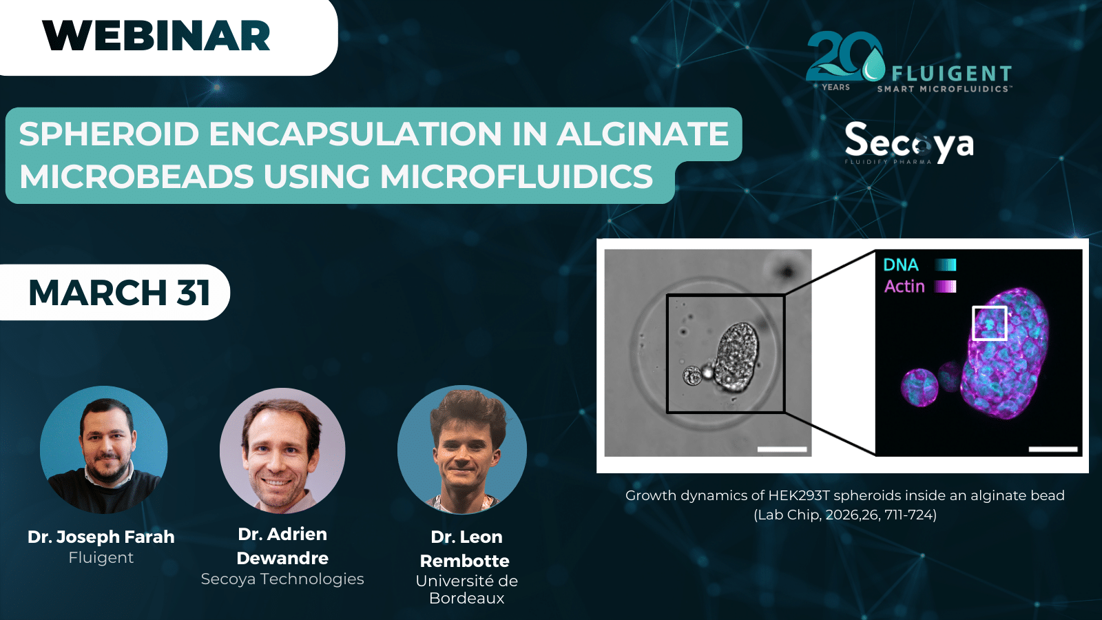
Spheroid encapsulation enables the development of controlled 3D cell culture models for studying tissue growth. In this case study, a droplet microfluidics approach was used for microfluidic encapsulation of mammalian cells in alginate microbeads using the RayDrop encapsulation platform. The method relies on a double emulsion with an alginate core and a sacrificial oil shell that enables homogeneous hydrogel crosslinking. Encapsulated cells remain viable and proliferate to form multicellular spheroids within the elastic alginate matrix.
A Paper for Université de Bordeaux, Université Libre de Bruxelles & Secoya Technologies
This study is a collaboration between Université de Bordeaux, CNRS, Université Libre de Bruxelles, and Secoya Technologies, a spin-off developing lab-scale equipment for (bio)-pharmaceutical processes. Their RayDrop® microfluidic droplet generator integrates Secoya’s emulsification technology into an easy-to-use platform, enabling reproducible spheroid encapsulation and droplet generation for academic and R&D labs.
Next Webinar: Spheroid Encapsulation in Alginate Microbeads Using Microfluidics
Join this session to learn how microfluidics can enable monodispersed alginate microbeads, high cell viability, and controlled spheroid growth in 3D environments.
📅 Two live sessions available on March 31st :
• 10:00 AM CEST – Register here
• 4:00 PM CEST – Register here
What Challenges Limit Long-Term 3D Cell Culture?
Spheroid encapsulation is a key technique for generating reproducible 3D cell culture models, including organoids for regenerative medicine and multicellular spheroids (MCS) for oncology. Compared to traditional 2D culture, these models better reproduce tissue physiology, but their production has often relied on labor-intensive protocols, limiting scalability for high-throughput applications. Droplet microfluidics enables precise microfluidic encapsulation of cells within hydrogel droplets, supporting self-assembly into spheroids while maintaining controlled microenvironments.1–4
Hydrogel scaffolds, such as alginate, provide both mechanical support and biochemical cues essential for long-term MCS culture. Conventional methods for generating alginate microbeads often produce heterogeneous sizes and may compromise cell viability (Figure 1). Advanced strategies using double emulsions in microfluidic systems improve bead uniformity, yet controlling gentle gelation without affecting cells remains challenging.5
Monodisperse hydrogel microbeads with uniform size and structure are critical for high-quality spheroid encapsulation, enabling consistent nutrient diffusion and optimal cell growth. Integrating controlled microfluidic techniques with biocompatible hydrogels addresses the main limitations of conventional MCS production methods (Table 1).6,7

Figure 1: Illustration of current strategies to produce 3D spheroids (Journal of Drug Delivery Science and Technology 2024, 100, 106033).
Table 1: Comparison of some current strategies for cell encapsulation in alginate beads ( Lab Chip, 2026,26, 711-724).
| Reference | Emulsion Type | Gelation Mechanism | Bead Diameter [μm] | Cell Viability | MCS Formation | Special Remarks |
|---|---|---|---|---|---|---|
| Lian et al. 8 | Simple | CaCl₂ in oleic acid | 40–55 | NA | NA | Cell viability not discussed. Encapsulation of bacteria, not mammalian cells. Involves oil removal steps. |
| Kim 9 | Simple | CaCl₂ in oleic acid | 100 | >85% | Yes | Clogging issues. Involves oil removal steps. |
| Trivedi et al. 10 | Simple | Droplet merging | 1500 | 80% and 60% after 4 h and 9 h | No | Viability decreases with time after encapsulation. Large bead diameters. Involves oil removal steps. |
| Akbari & Pirbodaghi 11 | Simple | De-chelation CaCO₃ (acidification) | 26 | 85% | Yes | Cell viability varies with time (74% after 2 days, 84% after 6 days). Involves fluorinated oil removal steps. |
| Liao et al. 12 | Double | Ca²⁺ diffusion through an oil shell | 190–260 | 78.1% | No | Poor shape control. Doubtful mechanism of crosslinking. Viability measured only 2 h after encapsulation. Beads directly recovered in aqueous medium. |
| Kieda et al. 13 | Double | CaCl₂ in continuous phase | 209 | 48% after encapsulation; 95% after 2 days | Yes | All-aqueous microfluidics. Shell phase acts as shield to prevent device clogging. Large polydispersity (CV 13%), excellent bead sphericity, monodispersed. |
Aim of the Study: Sacrificial Oil-Shell Method for Spheroid Encapsulation
The aim of this work is to develop a robust and biocompatible approach for spheroid encapsulation suitable for mammalian cells in 3D cell culture systems.
A droplet microfluidics strategy, based on a sacrificial oil-shell, was optimized was optimized to generate homogeneous alginate microbeads through controlled calcium diffusion across a thin oil shell, enabling gentle and uniform hydrogel crosslinking (Figure 2).
This approach allows efficient microfluidic encapsulation while preserving cell viability and supporting the formation of multicellular spheroids. Notably, this work demonstrates for the first time the use of a non-embedded capillary geometry to produce monodisperse and structurally homogeneous alginate microbeads compatible with long-term spheroid culture.

Materials and Methods: How to Set Up a Microfluidic Platform for Spheroid Encapsulation
Mammalian HEK293T cells were used for 3D cell culture experiments. Cells were prepared in a sterile alginate solution to enable their encapsulation within hydrogel microbeads.
Encapsulation was performed using the RayDrop (developed and manufactured by Secoya Technologies), a capillary-based droplet microfluidics device with a non-embedded co-focusing geometry. The system features three inlets for the core (cell-alginate solution), shell (oleic acid), and continuous phase (aqueous solution with surfactant) and a single outlet for microbead collection (Figure 3).
Precise flow control of all phases was achieved using the Flow EZ pressure controller, allowing stable, pressure-driven injection and reproducible droplet formation. In-line filters were placed on all phases to remove particulates and prevent device clogging. A sample injector with a loop was used for the cell-containing core to prevent sedimentation and ensure uniform loading (Figure 4).
Droplet generation was monitored using an optical camera, and the microfluidic setup was configured to maintain coaxial flow through the nozzle into the extraction capillary, ensuring proper formation of double-emulsion droplets. All tubing and components in contact with cells were sterilized prior to use.


Material Used for Spheroid Encapsulation


Proof of Concept: Chelate-Free Encapsulation of Mammalian Cells for Spheroid Formation
A- Cell Encapsulation in Alginate Microbeads
Mammalian HEK293T cells were encapsulated using the chelate-free CaCl₂ approach within alginate microbeads generated by droplet microfluidics. The process relies on calcium diffusion through a sacrificial oleic acid shell surrounding the alginate core (Figure 5).
Double emulsions were produced with the RayDrop platform, using coaxial flow: the alginate-cell solution forms the core, oleic acid forms the shell, and a surfactant-containing aqueous phase acts as the continuous phase. Controlled flow rates (core: 25 μL/min; shell: 15 μL/min; continuous: 300 μL/min) generate monodisperse droplets with diameters ranging from 200 to 400 μm.
Calcium ions diffuse from the collection bath through the oleic acid shell, inducing slow and uniform alginate gelation. After gelation, the oil shell detaches spontaneously, and the microbeads are recovered via gentle centrifugation, yielding structurally homogeneous, spherical beads ready for culture.

B- Spheroid Formation and Growth
Encapsulated HEK293T cells aggregate and form multicellular spheroids within the elastic alginate microbeads. Spheroid growth follows an exponential trend, consistent with the expected cell division times (~12–20 h). Small spheroids remain largely spherical, while larger ones show slight deviations in circularity due to mechanical constraints from the hydrogel (Figure 6).
The elastic alginate network promotes the formation of a supracellular F-actin shell at the spheroid boundary and limits coalescence between adjacent spheroids. This approach ensures high cell viability, reproducible spheroid formation, and controlled growth dynamics suitable for long-term 3D culture studies.

Conclusion
This case study presents a practical and reproducible approach for encapsulating mammalian cells in monodisperse, homogeneous alginate microbeads using the RayDrop platform combined with Flow EZ pressure controllers for precise flow regulation. Slow diffusion of Ca²⁺ through the oleic acid shell enables uniform gelation, while spontaneous oil detachment allows easy bead recovery.
This method maintains high cell viability, supports long-term spheroid formation, and can be easily implemented in standard biology labs without prior microfluidics expertise, offering a robust tool for studying tissue morphogenesis and 3D cell culture dynamics.
Read the full paper: Rembotte, L.; Cappello, J.; Dewandre, A.; Mettler, M.; Septavaux, J.; Nassoy, P.; Scheid, B. Sacrificial Oil Shell Method for the Generation of Alginate Microbeads Adapted to Multicellular Spheroid Culture. Lab on a Chip 2026.
Related Resources
-
Expert Reviews: Basics of Microfluidics The Role of Microfluidics in Advanced Organoid Modeling: from Static to Dynamic Read more
-
Expert Reviews: Basics of Microfluidics Microgel Crosslinking: Materials, Methods, and Emerging Applications Read more
-
Expert Reviews: Basics of Microfluidics 10 Tips for Reliable Droplet Generation Read more
-
Microfluidic Application Notes A quick and efficient double encapsulation method for FACS-based droplet sorting Read more
-
Microfluidics White Papers Double emulsion for the generation of microcapsules – a Review Read more
-
Expert Reviews: Basics of Microfluidics Flow control for droplet generation using syringe pumps and pressure-based flow controllers Read more
Webinar Replay of Interest


Related Solutions

References
(1) Rembotte, L.; Cappello, J.; Dewandre, A.; Mettler, M.; Septavaux, J.; Nassoy, P.; Scheid, B. Sacrificial Oil Shell Method for the Generation of Alginate Microbeads Adapted to Multicellular Spheroid Culture. Lab on a Chip 2026.
(2) Lampart, F. L.; Iber, D.; Doumpas, N. Organoids in High-Throughput and High-Content Screenings. Front. Chem. Eng. 2023, 5. https://doi.org/10.3389/fceng.2023.1120348.
(3) Jensen, C.; Teng, Y. Is It Time to Start Transitioning From 2D to 3D Cell Culture? Front. Mol. Biosci. 2020, 7. https://doi.org/10.3389/fmolb.2020.00033.
(4) Fevre, R.; Mary, G.; Vertti-Quintero, N.; Durand, A.; Tomasi, R. F.-X.; Del Nery, E.; Baroud, C. N. Combinatorial Drug Screening on 3D Ewing Sarcoma Spheroids Using Droplet-Based Microfluidics. Iscience 2023, 26 (5).
(5) Chae, S.; Hong, J.; Hwangbo, H.; Kim, G. The Utility of Biomedical Scaffolds Laden with Spheroids in Various Tissue Engineering Applications. Theranostics 2021, 11 (14), 6818.
(6) Gadziński, P.; Froelich, A.; Jadach, B.; Wojtyłko, M.; Tatarek, A.; Białek, A.; Krysztofiak, J.; Gackowski, M.; Otto, F.; Osmałek, T. Ionotropic Gelation and Chemical Crosslinking as Methods for Fabrication of Modified-Release Gellan Gum-Based Drug Delivery Systems. Pharmaceutics 2022, 15 (1), 108. https://doi.org/10.3390/pharmaceutics15010108.
(7) Arora, S.; Singh, S.; Mittal, A.; Desai, N.; Khatri, D. K.; Gugulothu, D.; Lather, V.; Pandita, D.; Vora, L. K. Spheroids in Cancer Research: Recent Advances and Opportunities. Journal of Drug Delivery Science and Technology 2024, 100, 106033. https://doi.org/10.1016/j.jddst.2024.106033.
(8) Lian, M.; Collier, C. P.; Doktycz, M. J.; Retterer, S. T. Monodisperse Alginate Microgel Formation in a Three-Dimensional Microfluidic Droplet Generator. Biomicrofluidics 2012, 6 (4).
(9) Kim, C. Droplet-Based Microfluidics for Making Uniform-Sized Cellular Spheroids in Alginate Beads with the Regulation of Encapsulated Cell Number. BioChip J 2015, 9 (2), 105–113. https://doi.org/10.1007/s13206-015-9203-6.
(10) Trivedi, V.; Ereifej, E. S.; Doshi, A.; Sehgal, P.; VandeVord, P. J.; Basu, A. S. Microfluidic Encapsulation of Cells in Alginate Capsules for High Throughput Screening. In 2009 Annual International Conference of the IEEE Engineering in Medicine and Biology Society; IEEE, 2009; pp 7037–7040.
(11) Akbari, S.; Pirbodaghi, T. Microfluidic Encapsulation of Cells in Alginate Particles via an Improved Internal Gelation Approach. Microfluid Nanofluid 2014, 16 (4), 773–777. https://doi.org/10.1007/s10404-013-1264-z.
(12) Liao, Q.-Q.; Zhao, S.-K.; Cai, B.; He, R.-X.; Rao, L.; Wu, Y.; Guo, S.-S.; Liu, Q.-Y.; Liu, W.; Zhao, X.-Z. Biocompatible Fabrication of Cell-Laden Calcium Alginate Microbeads Using Microfluidic Double Flow-Focusing Device. Sensors and Actuators A: Physical 2018, 279, 313–320.
(13) Kieda, J.; Appak-Baskoy, S.; Jeyhani, M.; Navi, M.; Chan, K. W. Y.; Tsai, S. S. H. Microfluidically-Generated Encapsulated Spheroids (μ-GELS): An All-Aqueous Droplet Microfluidics Platform for Multicellular Spheroids Generation. ACS Biomater. Sci. Eng. 2023, 9 (2), 1043–1052. https://doi.org/10.1021/acsbiomaterials.2c00963.