Liver-on-a-Chip: Models, Drug Metabolism and Applications
Liver-on-a-chip systems maintain hepatic cells under controlled perfusion and reproduce selected features of the liver microenvironment that are difficult to sustain in static culture. They are used to study hepatotoxicity, drug-induced liver injury (DILI), drug metabolism, ADME, metabolic liver disease and inter-organ pharmacology.
What Is a Liver-on-a-Chip?
A liver-on-a-chip is a microfluidic culture device in which hepatocytes are maintained in perfused channels or chambers, alone or with non-parenchymal cells.
The organ-on-chip approach combines microfabricated culture compartments with controlled fluid handling to reproduce selected tissue-level functions and microenvironmental cues rather than attempting to recreate the whole organ [1].
How does a liver-on-a-chip work?
Perfusion renews oxygen and nutrients, removes soluble waste, enables controlled dosing and serial sampling, and changes transport conditions around the cells. Device material is an equally important design variable: adsorption or absorption of test compounds can change the concentration that reaches the cells.
What chip architectures are used?
Architectures vary with the experimental question. Membrane devices can separate hepatic and endothelial compartments, while scaffold- and spheroid-based devices provide a 3D environment that supports cell-cell and cell-matrix interactions.
In the CNRS/UTC hydroscaffold model, HepG2/C3A cells were perfused at 10 µL/min for 21 days and formed large 3D spheroids while maintaining high viability and albumin production [2]. This experimental architecture is shown in Figure 1.

Figure 1. CNRS/UTC liver-on-a-chip model integrating a 3D hydroscaffold, dynamic culture and hepatic functional readouts. Source: CNRS/UTC case study; experimental work: Messelmani et al., Bioengineering (2022) [2].
Which hepatic cell sources are available?
Primary human hepatocytes remain an important reference for metabolic competence, although availability, donor variability and phenotype stability can limit routine use. HepaRG, HepG2/C3A and iPSC-derived hepatocyte-like cells are therefore also used, depending on the endpoint.
Differentiated HepaRG cells express the major cytochrome P450s, phase 2 conjugating enzymes and nuclear receptors at levels comparable to cultured primary human hepatocytes [13].
Why does shear stress matter in a liver-on-a-chip?
Shear stress is a further design parameter with a narrow working window (Figure 2). Hepatic sinusoids experience fluid shear well below that of most other capillary beds, and values reported for the sinusoidal range are of the order of 0.1-0.5 dyn/cm² against roughly 15 dyn/cm² elsewhere [3]. Flow rate, channel geometry and cell position therefore have to be reported together. Wider context on the format is available in the Fluigent guide to organs-on-chips.

Figure 2. Schematic cells under static conditions (without shear stress) and under fluid flow generating shear stress.
Recreating Hepatic Architecture and Zonation on Chip
Liver function is spatially organized along the portal-central axis. Periportal hepatocytes are exposed to relatively higher oxygen and preferentially support pathways including oxidative metabolism, gluconeogenesis and fatty-acid oxidation. Pericentral hepatocytes experience lower oxygen and are enriched in glycolytic, lipogenic and several xenobiotic-metabolism pathways [4,5].
Can a chip reproduce hepatic zonation?
Microfluidic systems can generate controlled gradients that reproduce selected aspects of this zonation. Tonon and colleagues designed a device that imposed a stable oxygen gradient independently of medium perfusion. The chip design and oxygen-gradient validation are shown in Figure 3 [5].

Figure 3. Microfluidic chip design and oxygen-gradient validation. From Tonon et al., Scientific Reports 9, 13557 (2019) [5].
The gradient produced zonated glycogen storage, higher GLUL expression at low oxygen, higher CPS1 expression at high oxygen and increased CYP3A4 toward the low-oxygen side [5].
This is more precise than stating that a chip reproduces the entire hepatic lobule: oxygen is a major cue, but it is not the only determinant of zonation.
The corresponding CYP3A4 and glycogen readouts are shown in Figure 4.

Figure 4. CYP3A4 expression and glycogen-storage readouts across an imposed oxygen gradient. From Tonon et al., Scientific Reports (2019) [5].
Which cells contribute to hepatic function?
Cellular composition adds another level of physiology. Liver sinusoidal endothelial cells contribute filtration, endocytosis and immune surveillance; Kupffer cells provide resident innate immune functions; and hepatic stellate cells are central to fibrogenic remodeling [6].
Flow rate, channel geometry and cell position should therefore be reported together, because the shear stress transmitted to cells depends on the complete fluidic architecture.
How are drug-metabolising enzymes distributed?
Enzyme distribution follows the same axis. Quantitative cross-species imaging places CYP2E1 across the whole pericentral region with extension into the midzone, and CYP3A4 in zone 3 extending into zone 2 in human liver, where the periportal-to-perivenous gradient was steeper than in the other species examined [4].
Because the same isoforms bioactivate several compounds, the zone a model reproduces influences which toxicities it can detect.
Liver-on-a-Chip for Drug Metabolism and DILI Studies
The liver is a major site of xenobiotic metabolism, so hepatic microphysiological systems can address complementary questions: whether a compound injures liver cells, how functional markers change over time, how metabolites are generated, how rapidly a parent compound disappears and how inflammatory or disease states modify those responses [14].
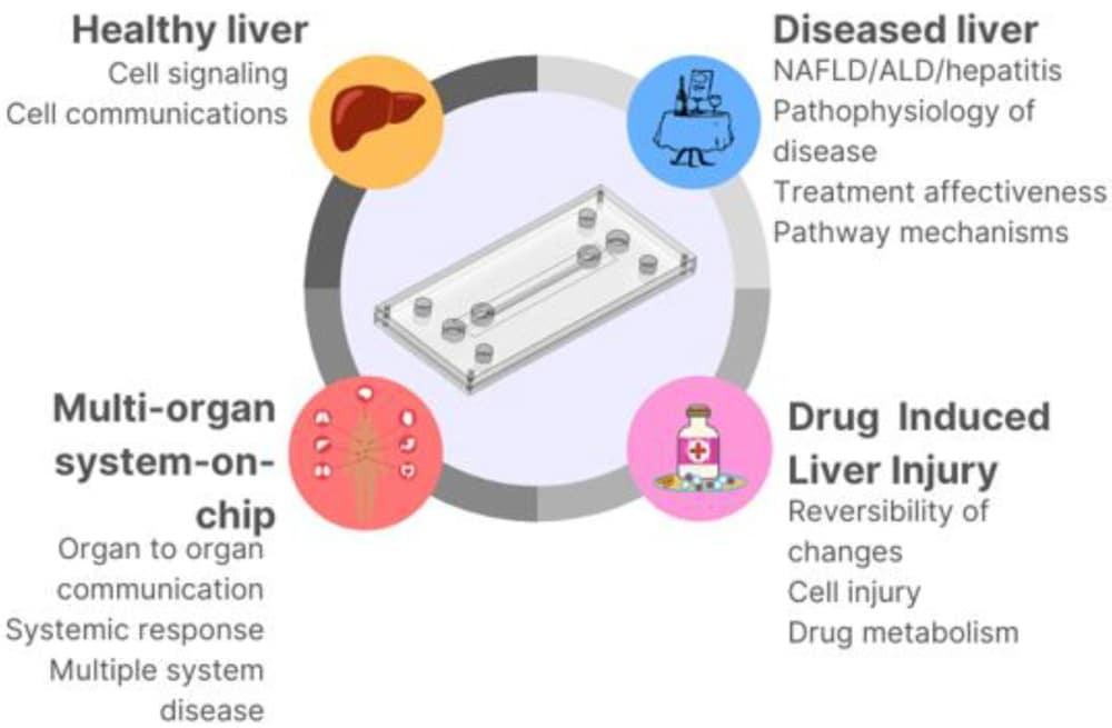
Figure 5. The use of microfluidic devices in different aspects of healthy and liver disease modeling. From Kanabekova et al., Micromachines (2019).
Hepatotoxicity screening
Hepatotoxicity studies commonly combine viability with functional readouts such as albumin secretion, urea production and cytochrome P450 activity.
Perfusion makes repeat dosing and serial medium sampling easier over extended experiments, so the analysis yields a time course rather than a single endpoint. In regulatory toxicology this complements, rather than replaces, conventional animal testing.
Drug-induced liver injury (DILI)
Drug-induced liver injury remains a major safety concern. In a systematic review of 462 medicinal products withdrawn worldwide between 1953 and 2013, hepatotoxicity was the most frequently reported adverse reaction leading to withdrawal: 81 cases, or 18% [7].
Liver-on-chip models can add time-resolved and mechanistic information, particularly when co-cultures include immune or stellate-cell components. Poor correspondence between traditional animal models and human clinical outcomes is the stated reason preclinical strategy has moved toward human in vitro systems with multi-parametric endpoints [8].
Idiosyncratic DILI is the harder case, since it depends partly on immune and inflammatory mechanisms that hepatocyte monoculture does not contain. Adding Kupffer cells introduces resident innate immune activity and stellate cells report fibrogenic response, which is why co-culture design and cell sourcing are reported alongside the toxicity endpoint. How liver models sit within the wider preclinical pipeline is covered in the organ-on-chip in drug development review.
ADME and clearance
Sequential sampling enables metabolite identification and compound-depletion experiments. Interpretation requires more than nominal dose: inlet concentration, flow rate, residence time, cell number, system volume, recirculation and nonspecific material loss all influence the exposure experienced by cells.
Material selection can be decisive for quantitative pharmacology. In a 2025 study, imipramine fell from 100 µM to 0.0384 µM after 24 h in PDMS devices, compared with 31.5 µM in cyclic olefin copolymer under the tested static conditions [9].
Steatosis and NASH models
Metabolic liver disease can be modeled by changing the biochemical environment while retaining a controlled 3D culture architecture.
NAFLD and NASH remain the terms used across most of the published chip and spheroid literature, including the studies cited in this section, so they are retained here.
Current clinical nomenclature replaces them with metabolic dysfunction-associated steatotic liver disease (MASLD) and metabolic dysfunction-associated steatohepatitis (MASH) respectively [15]; where a cited study used the older terms, those are kept in the description of that study.
Why Recirculation Matters for Metabolism Studies
How does a recirculation loop work?
Recirculation returns medium from the outlet side of the system to the inlet rather than continuously discarding it.
This can allow secreted factors, metabolites and drug products to accumulate, which is useful for metabolite-transfer studies, long exposures and experiments where the concentration-time profile itself is an endpoint. Recirculating drugs and their metabolites is what makes longer-term pharmacokinetic measurement on chip possible [16].
What changes in a closed loop?
Closed-loop operation also changes system pharmacokinetics. Parent drug concentration can decrease as the liver model clears the compound while metabolites accumulate.
Recirculation should not automatically be considered “more physiological”, however: loop volume, medium exchange, binding to tubing or chip materials, evaporation and waste accumulation can all influence the result.
Published design guidance for recirculating systems is to avoid drug-permeable materials, keep medium volumes low so that secreted factors are not diluted, sample the medium throughout the run, and pair the experiment with a mass-balance or pharmacokinetic model for interpretation [16].
Three classes of experiment depend on it: prodrug studies, where the active metabolite has to persist; autocrine and paracrine signalling, where secreted factors must be retained; and chronic exposure, where a defined circulating dose is needed rather than a continuously refreshed one. The fluidic design considerations are set out in the importance of recirculation in microfluidic systems review.
Fluigent Microfluidic Recirculation Pack
Fluigent’s pressure-driven Recirculation Pack combines flow control, sensing and automated switching for stable unidirectional recirculation. See the Microfluidic Recirculation Pack. The recirculation configuration is shown in Figure 6.
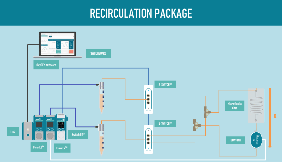
Figure 6. Fluigent microfluidic recirculation setup.
Multi-Organ Models: Liver-Kidney and Beyond
Why couple a liver model to another organ?
Multi-organ systems connect a liver compartment to another tissue so that liver-generated metabolites can affect a downstream organ. The technology couples two or more cell types under shared perfusion. This is particularly relevant when toxicity or clearance depends on hepatic biotransformation followed by renal, cardiac or other tissue exposure. An example of liver-kidney model is presented in figure 7 (from Fluigent/University of Limoges application note).
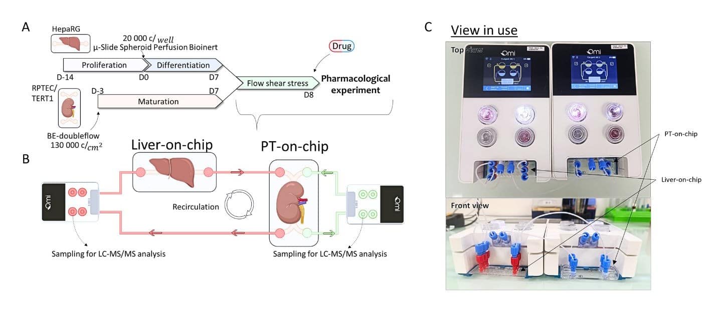
Figure 7. Liver-Kidney Dual OOC setup using the Omi Dual platform. Source: Fluigent / University of Limoges application note.
What did the liver-kidney study measure?
In the Fluigent/University of Limoges application, HepaRG liver spheroids and RPTEC/TERT1 proximal-tubule cells were coupled under automated recirculation. The renal circuit was operated at 10 µL/min and the hepatic circuit at 20 µL/min for 48 hours.
Tacrolimus decreased rapidly in the hepatic compartment and was later detected in the urinary compartment; DM-tacrolimus was detected in both compartments.
The experiment illustrates both the biological value and the engineering constraints of multi-organ coupling: medium compatibility, compartment volumes, transport rates and scaling assumptions must all be defined.
The corresponding tacrolimus and DM-tacrolimus measurements are shown in Figure 8.
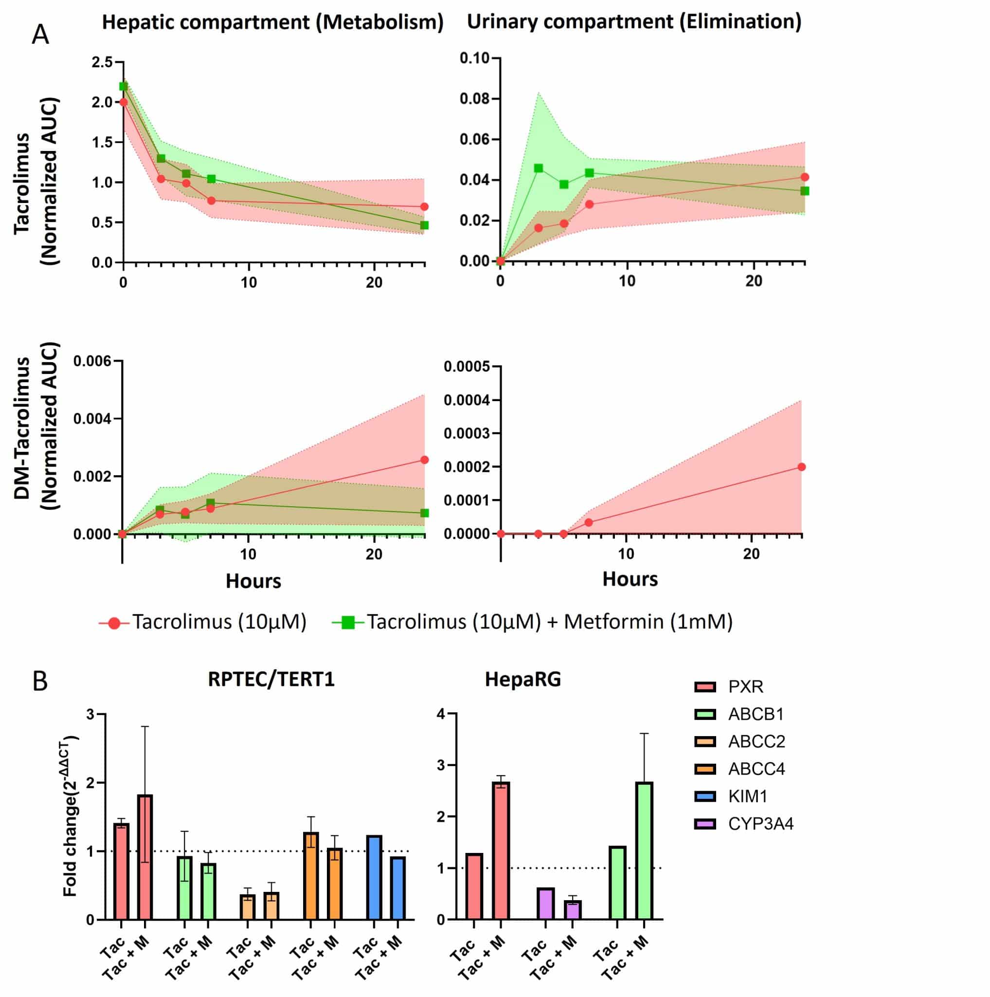
Figure 8. Tacrolimus and DM-tacrolimus measurements in the dual-organ model. Source: Fluigent / University of Limoges application note.
In the reported data, tacrolimus fell quickly in the hepatic compartment and stabilised after roughly eight hours, while the urinary compartment peaked later at about a tenth of the hepatic concentration, consistent with biliary-dominant clearance. Method and dataset are given in the liver-kidney organ-on-chip application note, with a recorded webinar on the model.
A wider approach has been applied at larger scale in a recent paper Ronaldson-Bouchar et al . (Nature Biomedical Engineering, 2022). Recirculating vascular flow has been used to link matured human heart, liver, bone and skin tissues behind selectively permeable endothelial barriers, maintaining their phenotypes over four weeks of culture and reproducing the pharmacokinetic and pharmacodynamic profile of doxorubicin, with a compartmental mass-balance model used to interpret the concentration data [17].
Case Study: A Liver-on-a-Chip Model (CNRS)
What did this liver-on-a-chip model show?
Messelmani and colleagues integrated a hyaluronic-acid hydroscaffold containing liver extracellular-matrix components and RGDS adhesion sites into a microfluidic liver model [2].
HepG2/C3A cells were perfused at 10 µL/min for 21 days, self-organized into 3D spheroids and maintained high viability and albumin production compared with static controls.
Why is pressure monitoring useful?
The study also shows why pressure monitoring can be informative in long-term perfusion. At the target flow rate, pressure was approximately stable during early culture and then increased when proliferating aggregates occupied more of the chamber.
In this article data, pronounced obstruction at day 21 was accompanied by a large pressure rise.
Pressure therefore acted as an indirect readout of increasing hydraulic resistance and impending clogging rather than relying on flow-rate information alone. The associated pressure evolution is shown in Figure 9.
Pressure remained close to 60 mbar through day 11 as the spheroids grew, then rose sharply once aggregates occupied enough of the chamber to restrict the flow path. Full protocol, figures and data are in the liver-on-chip case study.


Figure 9. Pressure evolution during the CNRS/UTC liver-on-a-chip culture. Source: Messelmani et al., Bioengineering (2022) [2].
Equipment for Liver-on-a-Chip Experiments
What equipment does a liver-on-a-chip require?
A typical liver-on-a-chip setup combines a stable flow source, a chip or culture chamber, tubing and connectors matched to the flow range, and where required flow sensing, switching, sampling or recirculation.
Hydraulic resistance depends on both chip geometry and the internal diameter and length of tubing, so the full fluidic layout should be kept consistent across replicates.
| Need | Typical Fluigent option |
|---|---|
| Modular pressure-driven perfusion | MFCS EZ or Flow EZ pressure controller, optionally paired with Flow Unit sensing and feedback control. |
| Integrated organ-on-chip automation | Omi for programmable perfusion, recirculation, sampling and compound delivery. |
| Configured perfusion workflow | Organ on Chip Perfusion Pack with pressure-driven control, sensing and compatible connections. |
| Closed-loop recirculation | Microfluidic Recirculation Pack with automated switching and sensing. |
Why choose pressure-driven flow control?
Pressure-driven control is particularly useful when experiments require responsive flow adjustment, long-term perfusion and integration with real-time flow sensing. Figure 10 shows the components of the Microfluidic Recirculation Pack.
For a modular setup the Flow EZ pressure controller provides pulseless pressure-driven flow; the cell perfusion package supplies a configured single-pass workflow, and the automated organ-on-chip platform integrates perfusion, recirculation and sampling for multi-chip operation.


Figure 9. Pressure evolution during the CNRS/UTC liver-on-a-chip culture. Source: Messelmani et al., Bioengineering (2022) [2].
Frequently Asked Questions
What is a liver-on-a-chip used for?
Liver-on-a-chip systems are used in preclinical research for hepatotoxicity and DILI, drug metabolism and compound-depletion experiments, ADME studies and disease modeling. They can also provide the hepatic compartment of a multi-organ system in which liver-generated metabolites are transferred to another tissue.
How does liver-on-a-chip compare with organoids for DILI?
Organoids provide self-organized 3D architecture, whereas organ-on-chip devices add externally controlled perfusion, gradients, dosing and serial sampling. The approaches can be combined by perfusing spheroids or organoids inside microfluidic devices [12].
Why is recirculation important in liver-on-a-chip?
Recirculation can prevent metabolites and secreted factors from being continuously washed away, allowing them to accumulate and influence the concentration-time profile. It is useful for long exposures, metabolite-transfer studies and some prodrug or clearance experiments, but loop volume, material binding, waste accumulation and medium-exchange schedules must be controlled.
Which cells are used in liver-on-a-chip models?
Primary human hepatocytes, HepaRG cells, HepG2/C3A cells and iPSC-derived hepatocyte-like cells are all used, depending on the question. These cell types differ in metabolic competence and availability. Endothelial cells, Kupffer cells and hepatic stellate cells can be incorporated to add vascular, inflammatory or fibrotic functions.
Related Expertises
-
Expert Reviews: Basics of Microfluidics Organ-on-a-Chip vs Organoids: Differences, Strengths and When to Combine Them Read more
-
Expert Reviews: Basics of Microfluidics Organ-on-Chip in Drug Development: A Human-Relevant Alternative to Animal Testing Read more
-
Expert Reviews: Basics of Microfluidics The Importance of Recirculation in Microfluidic Systems: Principles and Applications Read more
-
Expert Reviews: Basics of Microfluidics 5 Key Tips for Starting Organ-on-Chip Models Read more
-
Microfluidic Application Notes Liver–Kidney Organ-On-Chip Model using the Omi™ Dual Platform Read more
-
Microfluidics Case Studies CNRS/UTC: study of a liver-on-a-chip model Read more
-
Microfluidics White Papers A review of Organ on Chip Technology – A White Paper Read more
References
- Bhatia SN, Ingber DE. Microfluidic organs-on-chips. Nature Biotechnology. 2014;32:760-772. https://doi.org/10.1038/nbt.2989
- Messelmani T, Le Goff A, Souguir Z, et al. Development of Liver-on-Chip Integrating a Hydroscaffold Mimicking the Liver’s Extracellular Matrix. Bioengineering. 2022;9:443. CC BY 4.0. https://doi.org/10.3390/bioengineering9090443
- Rashidi H, Alhaque S, Szkolnicka D, Flint O, Hay DC. Fluid shear stress modulation of hepatocyte-like cell function. Archives of Toxicology. 2016;90:1757-1761. https://doi.org/10.1007/s00204-016-1689-8
- Albadry M, Küttner J, Grzegorzewski J, et al. Cross-species variability in lobular geometry and cytochrome P450 hepatic zonation. Frontiers in Pharmacology. 2024;15:1404938. CC BY.
- Tonon F, Giobbe GG, Zambon A, et al. In vitro metabolic zonation through oxygen gradient on a chip. Scientific Reports. 2019;9:13557. CC BY 4.0. https://doi.org/10.1038/s41598-019-49412-6
- Shetty S, Lalor PF, Adams DH. Liver sinusoidal endothelial cells – gatekeepers of hepatic immunity. Nature Reviews Gastroenterology & Hepatology. 2018;15:555-567.
- Onakpoya IJ, Heneghan CJ, Aronson JK. Post-marketing withdrawal of 462 medicinal products because of adverse drug reactions. BMC Medicine. 2016;14:10. CC BY. https://doi.org/10.1186/s12916-016-0553-2
- Walker PA, Ryder S, Lavado A, Dilworth C, Riley RJ. Strategies to minimise the risk of human drug-induced liver injury. Archives of Toxicology. 2020;94:2559-2585. CC BY 4.0.
- Grindulis K, Matusevica NG, Kozlova V, et al. Sorption and release of small molecules in PDMS and COC for Organs on chip. Scientific Reports. 2025;15:14012. CC BY-NC-ND 4.0. https://doi.org/10.1038/s41598-025-97111-2
- Bulutoglu B, Rey-Bedón C, Kang YBA, et al. A microfluidic patterned model of non-alcoholic fatty liver disease: applications to disease progression and zonation. Lab on a Chip. 2019;19:3022-3031.
- Pingitore P, Sasidharan K, Ekstrand M, et al. Human Multilineage 3D Spheroids as a Model of Liver Steatosis and Fibrosis. International Journal of Molecular Sciences. 2019;20:1629. CC BY 4.0. https://doi.org/10.3390/ijms20071629
- Zhao Y, Landau S, Okhovatian S, et al. Integrating organoids and organ-on-a-chip devices. Nature Reviews Bioengineering. 2024;2:588-608.
- Aninat C, Piton A, Glaise D, et al. Expression of cytochromes P450, conjugating enzymes and nuclear receptors in human hepatoma HepaRG cells. Drug Metabolism and Disposition. 2006;34:75-83.
- Frojdenfal S, Zuchowska A. Advanced Liver-on-a-Chip Model for Evaluating Drug Metabolism and Hepatotoxicity. Biosensors. 2024;14:435. CC BY 4.0. https://doi.org/10.3390/bios14090435
- AASLD. New MASLD nomenclature / Steatotic Liver Disease terminology, announced 2023. https://www.aasld.org/new-masld-nomenclature
- Ronaldson-Bouchard K, Vunjak-Novakovic G. Organs-on-a-chip: a fast track for engineered human tissues in drug development. Cell Stem Cell. 2018;22(3):310-324. https://doi.org/10.1016/j.stem.2018.02.011
- Ronaldson-Bouchard K, Teles D, Yeager K, et al. A multi-organ chip with matured tissue niches linked by vascular flow. Nature Biomedical Engineering. 2022;6(4):351-371. https://doi.org/10.1038/s41551-022-00882-6


