Organ-on-a-Chip vs Organoids: Differences, Strengths and When to Combine Them
Organ-on-a-chip vs organoid is a recurring question in advanced in vitro modeling, and the two are complementary rather than competing. Organoids derive tissue architecture from intrinsic developmental programs; organ-on-a-chip devices impose geometry and apply defined convective and mechanical boundary conditions. This review compares both classes across architecture, mass transport, throughput and readouts, situates them among microphysiological systems, and examines the hybrid configuration in which organoids are cultured under controlled perfusion.
Key Takeaways
- Organoids self-organise from stem cells into tissue-like structures of high biological complexity, but are diffusion-limited and unperfused.
- Organ-on-a-chip devices impose engineered geometry, convective transport and mechanical loading, at the cost of spontaneous morphogenesis.
- The distinction is transport, not biology. Organoids rely on diffusion; chips add convection.
- Organoid-on-a-chip combines both: self-organized tissue under defined flow.
- Select by measurable variable, not technology — throughput, exposure control and mechanotransduction each favour a different model.
What Is an Organoid?
Organoids are 3D, self-organized, stem-cell-derived structures that arise from intrinsic developmental programs. They are generated from induced pluripotent stem cells (iPSCs), embryonic stem cells, or adult stem cells, typically embedded in a basement-membrane extract that supplies laminin and collagen IV. Given appropriate morphogen gradients, the cells establish apico-basal polarity and tissue-specific architecture — crypt-villus axes in intestinal organoids, cortical layering in brain organoids.
Organoids are frequently confused with spheroids and tumoroids, which occupy different positions on the complexity spectrum:
- Spheroids are simple 3D aggregates of one or more cell types, self-assembled under low-adhesion conditions such as hanging-drop or ultra-low attachment plates. They lack defined architecture and are used in medium- to high-throughput screening.
- Tumoroids are patient-derived tumor organoids generated from primary tumor tissue. They retain the parent tumor’s mutational profile and intratumoral heterogeneity, supporting precision oncology [1-3].
Culture format constrains the result: hydrogel micro-bead encapsulation narrows the size distribution relative to bulk gel. See also organoid modeling from static to dynamic culture.
What Is an Organ-on-a-Chip?
Organ-on-a-chip devices are microengineered systems combining human cells, controlled microfluidic flow and mechanical actuation to reproduce organ-level structure and function. Channel dimensions are typically 10²–10³ µm, giving Reynolds numbers well below unity: flow is laminar, inertial effects are negligible, and transverse mixing occurs by diffusion alone, so concentration fields are deterministic and calculable.
Architecture is imposed rather than emergent. Channel geometry, membrane pore size and co-culture layout are fixed by fabrication, which renders tissue-tissue interfaces — epithelium apposed to endothelium across a porous membrane — reproducible between devices. Convective transport also decouples nutrient supply from diffusion path length, sustaining tissue beyond the ~100–200 µm oxygen diffusion limit of static 3D culture [4]. For a full technical treatment, see the Fluigent white paper, A Guide to Organs-on-Chips.
Where Microphysiological Systems Fit
Microphysiological systems (MPS) is a collective term for in vitro models reproducing human physiological function at tissue, organ or multi-organ scale. MPS is the superset, not a third option: organoids and organ-on-a-chip devices are both MPS [5-6]. The distinction matters for regulatory submissions, where FDA and EMA guidance addresses MPS and New Approach Methodologies, not named technologies [7].
MPS include organ-on-chip systems, organoids, perfused tumoroids or spheroids, bioprinted microtissues, transwell co-cultures, and multi-organ “body-on-chip” platforms.
From spheroids to organ-on-a-chip: a complexity hierarchy
Advanced in vitro models span a continuum from unstructured 3D aggregates to perfused, mechanically actuated devices, illustrated in Figure 1:
Spheroids → Tumoroids → Organoids → Organ-on-Chip → Full Microphysiological Systems
Progression along this axis increases the number of physiological variables reproduced and lowers throughput per unit cost. That trade-off, not intrinsic superiority, governs model selection.

Figure 1: Complexity of in vitro systems
Organ-on-a-Chip vs Organoids: Side-by-Side Comparison
Organoids provide morphogenetic fidelity and donor genotype; chips provide convective transport, defined mechanical loading, low inter-device variance and optical access. The table 1 extends the four-way comparison with vascularization, throughput, readouts and cost.
| Feature | Spheroids | Tumoroids | Organoids | Organ-on-chip |
|---|---|---|---|---|
| 3D structure | Simple sphere | Patient-derived structure | Organ-like | Engineered microenvironment |
| Cell source | Cell lines / mixed | Primary tumor | Stem cells | Primary or iPSC-derived |
| Architecture | Minimal | Tumor-specific | Tissue-specific, self-organized | Device-defined |
| Perfusion | None | None | None (unless hybrid) | Controlled |
| Vascularization | None | None | Rare, non-perfused | Perfusable endothelial channels |
| Mechanical forces | None | None | None | Physiological, tunable |
| Shear stress | None | None | Minimal | Fully tunable |
| Throughput | High | Medium | Medium | Low to medium |
| Readouts | Endpoint assays | Endpoint, patient-specific | Endpoint, imaging-limited | Live imaging, effluent sampling, biosensors |
| Reproducibility | Good | Medium | Medium–high | High |
| Personalisation | Low | Very high | High | Medium–high |
| Relative cost per data point | Low | Medium | Medium | High |
| Typical use | Medium-throughput screening | Patient-specific response | Disease-specific mechanism | Mechanistic PK/PD |
Tissue architecture and self-organisation
Organoids generate architecture through morphogen-driven patterning; chips receive it from fabrication. Stem cells establish crypt-villus axes, cortical plates or nephron segments without external templating, producing cell-type diversity that no device geometry specifies.
The cost is variance: organoid diameter, shape and composition disperse within and between batches, because self-organization amplifies small differences in seeding density and matrix lot. Device-defined culture suppresses that dispersion at the expense of emergent structure.
Vascularisation and perfusion
This is the sharpest mechanistic distinction. Organoids have neither vasculature nor perfusion, so oxygen and nutrient delivery is governed by Fickian diffusion, and consumption exceeds supply beyond a few hundred micrometres — producing a hypoxic, ultimately necrotic core. Chips replace diffusion with convection through defined channels.
Flow is also a signal, not only a supply route. Endothelial cells transduce wall shear stress of order 0.1–2 Pa into alignment, junctional remodelling and altered gene expression; epithelial cells respond with changes in differentiation and transporter abundance. Reproducing these responses requires low-pulsatility flow, which is why pressure-driven control such as the Flow EZ is used in place of stepper-motor syringe drives, whose displacement ripple imposes an unintended oscillatory shear component.
Control over the microenvironment
Chips decouple parameters confounded in static culture. Oxygen tension, cytokine gradients, drug concentration and mechanical strain are each variable while the others are held constant — a precondition for mechanistic dose-response work.
In static organoid culture these follow from the medium-exchange schedule and construct geometry. Gradients form, but as uncontrolled consequences of consumption and diffusion rather than as set variables, which forecloses quantitative exposure-response analysis.
Throughput and reproducibility
Organoids scale in number; chips scale in precision. Hundreds of organoids occupy one plate, whereas each chip requires priming, bubble-free connection and flow monitoring, limiting parallelism.
Reproducibility inverts the ranking. Inter-device variation is lower in chips because geometry and flow rate are instrument-set rather than emergent, while organoid heterogeneity persists within a single batch. Parallel perfusion manifolds narrow the throughput gap by driving multiple chips from one pressure source.
Readouts and imaging
Chip geometry is compatible with live microscopy; bulk-gel organoid culture is not. A thin, optically flat, refractive-index-matched floor sits within the working distance of high-numerical-aperture objectives, permitting time-lapse acquisition across the experiment, while effluent is sampled serially for metabolites, cytokines or injury markers. Transepithelial electrical resistance is measurable continuously via integrated electrodes.
Organoids embedded in bulk hydrogel scatter light, sit at variable and often excessive focal depth, and generally require fixation and optical clearing, making each measurement terminal. Cell culture and organ-on-a-chip microscopy chips are designed for continuous observation.
Figure 2 summarizes the different key features of each models.

Figure 2: Differences between organoids, organ-on-a-chip and MPS
Strengths and Limitations of Each Model
Each model is limited by the property that defines it: self-organization in organoids, imposed control in chips. The strengths and limitations of the models are presented in table 2.
| Organoids | Organ-on-a-chip | ||
|---|---|---|---|
| Strenghts | Limitations | Strenghts | Limitations |
| Morphogenetic fidelity and tissue-specific cell-type diversity | Diffusion-limited; no vasculature or convective transport | Defined wall shear stress, strain and concentration boundary conditions | Lower parallelism and higher cost per data point |
| Donor-derived material retains genotype and heterogeneity | Batch dispersion in size and composition complicates quantification | Low inter-device variance and reproducible tissue interfaces | Requires fluidic handling expertise and instrumentation |
| Established protocols for intestinal, brain, liver, kidney and colorectal tissue | Undefined, lot-variable basement-membrane matrices in most protocols | Live imaging, serial effluent sampling, integrated electrical readouts | Sorption of small hydrophobic compounds into elastomeric device materials |
Table 2: Strenghts and limitations of organoids and organ-on-a-chip models
Organoid-on-a-Chip: Combining Both Approaches
Organoid-on-a-chip denotes culture of self-organized organoids within a perfused microfluidic device: the organoid supplies morphogenetic complexity, the chip supplies convective transport, defined gradients and mechanical loading. The configuration relieves the diffusion limit without substituting engineered geometry for developmental patterning [2].
Brain organoid-on-a-chip
Brain organoid-on-a-chip denotes cerebral organoids cultured under controlled perfusion. Central nervous system organoids are among the largest and longest-maintained models, and therefore the most severely transport-limited: as diameter grows, the unperfused core becomes hypoxic and necrotic, a recognised confound in studies of neurodevelopment and neurodegeneration [6].
Perfusing the chamber raises interior oxygen flux and permits controlled morphogen or compound delivery, while enabling time-resolved imaging and serial sampling of secreted markers over multi-week culture.
Kidney organoid and vasculature models
Kidney organoids form nephron-like segments — podocyte clusters, proximal tubule-like epithelium — but only rudimentary, non-perfused endothelial networks. Apposing an endothelial compartment under flow yields a perfusable model in which organoid and vasculature interact directly, and in which endothelial cells receive the shear stimulus absent in static culture.
A Fluigent customer study implemented this configuration using pressure-driven control to perfuse the endothelial channel alongside the organoid — see creating a kidney organoid-vasculature interaction model.

Figure 3: Schematic representation of the co-culture timeline of HUVECs and kidney organoids, indicating static culture, exercise of flow and addition and collection points of kidney organoids. From Menéndez et al., Scientific Report, 2022.
Adding controlled flow to organoids
Converting a static protocol to a perfused one requires a chamber accommodating a 3D construct, a flow source with low pulsatility and week-scale set-point stability, and parallel channels for replicates. Pressure-driven controllers settle rapidly after a set-point change and produce no displacement ripple, which matters when applying time-varying exposure profiles rather than constant concentrations.
Where concentration gradients are the independent variable, gradient-generating chips establish reproducible profiles across a 3D chamber by laminar stream splitting and recombination. For extended unattended culture, the Omi automated organ-on-a-chip platform integrates perfusion, recirculation and environmental control.
How Organoid and Organ-on-a-Chip Technologies Developed
The two technologies originate in different disciplines — developmental biology and microfabrication — and converged only recently, which is why their capabilities remain complementary.
From spheroids to self-organising organoids
Unstructured 3D aggregates preceded organoids by decades. The decisive result was that adult stem cells could establish tissue-like architecture without external templating; iPSC protocols then extended the approach to brain, kidney, liver and lung.
From microfabrication to perfused chips
Organ-on-a-chip derives from soft lithography rather than stem cell biology. Early devices established that mechanical and fluidic boundary conditions alone restore organ-level functions absent from static culture, defining the engineering principles still in use [4].
Applications in Disease Modeling, Toxicology and Personalised Medicine
Model selection follows the measured endpoint: screening favours organoids, exposure-resolved mechanism favours chips, systemic disposition favours multi-organ configurations. Therapy-response endpoints span both.
Cancer and tumor models
Tumoroids retain donor genotype and suit screening candidate therapies against a defined tumor. Adding perfusion and immune compartments reconstructs the tumor microenvironment — vasculature, stroma, infiltrating lymphocytes — required for immunotherapy endpoints static culture cannot support, since immune recruitment depends on flow-mediated adhesion cascades [5]. Colorectal and other epithelial carcinomas are the most developed organoid applications; vascularized tumor-on-chip the most developed hybrid.
Liver, lung and intestinal toxicology
Toxicology requires exposure control, where chips are strongest. Liver chips resolve drug-induced injury under clearance kinetics rather than fixed-concentration exposure; lung chips reproduce cyclic strain and an air-liquid interface; intestinal chips reproduce peristalsis-like deformation and barrier function measurable by transepithelial resistance. Linked multi-organ systems extend this to human-relevant ADME.
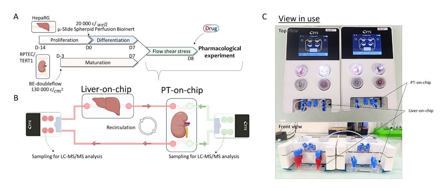
Figure 4: Overview of Liver-Kidney Dual OOC setup. In ollaboration with University of Limoges Pharmacology and Transplantation UMR 1248.
Patient-derived and personalization models
Personalized medicine requires donor-derived cells, which organoids supply most readily. Perfusion allows clinically realistic concentration-time profiles, rather than bolus exposure, to be applied to one patient’s tissue, and efficacy to be scored per donor.
How to Choose the Right Model for Your Study
Selection follows the variable to be measured:
- Screening across many compounds or donors — organoids or tumoroids; parallelism and genotype coverage outweigh transport fidelity.
- Dose-response, clearance or exposure-resolved mechanism — organ-on-a-chip; defined concentration-time profiles are required.
- Barrier integrity, shear-dependent phenotype, vascular biology or mechanical loading — organ-on-a-chip; these variables are undefined in static culture.
- Extended culture of large 3D tissue, or donor tissue under realistic dosing — organoid-on-a-chip.
A practical sequence is to establish the biology statically, then add flow: plate-based development is cheaper per iteration, and transfer to chip is tractable once organoid variance is characterized.
Equipment for Perfused Organoid and Organ-on-a-Chip Culture
Perfused organoid and organ-on-a-chip culture requires a flow source with low pulsatility and long-term set-point stability, chambers accommodating 3D constructs, and automation sufficient for multi-week operation. Pressure-driven control is preferred to syringe drives because it produces continuous, ripple-free flow and settles rapidly after a set-point change.
- Flow EZ — pressure-based flow controller, standalone or integrated, for defined shear boundary conditions.
- Omi, automated organ-on-a-chip platform — integrated perfusion, recirculation and environmental control for unattended long-term culture.
- Cell culture and organ-on-a-chip microscopy chips — optically compatible devices for combining organoids with controlled flow.
- Mammalian spheroid encapsulation case study — workflow for narrow-dispersion 3D constructs upstream of chip culture.
Conclusion
Organ-on-a-chip and organoids differ principally in how mass transport and mechanical boundary conditions are established: diffusive and emergent in one, convective and imposed in the other. Organoids provide morphogenetic fidelity and donor genotype; chips provide defined shear, strain and concentration fields with low inter-device variance. Combining them places self-organized tissue under specified transport conditions. Selection is therefore a question of which variables must be controlled and which measured — and, where neither suffices alone, of adding defined perfusion to the construct that already carries the required cells.
Frequently Asked Questions
What is the difference between an organoid and an organ-on-a-chip?
An organoid is a self-organized 3D tissue derived from stem cells, biologically complex but unperfused and diffusion-limited. An organ-on-a-chip is a microfluidic device imposing defined geometry, convective flow and mechanical loading on cultured cells. Organoids maximise morphogenetic fidelity; chips maximise control over transport and mechanical boundary conditions.
Can organoids be cultured on a chip?
Yes. Organoid-on-a-chip culture places organoids inside a perfused microfluidic device, retaining self-organized structure while adding convective nutrient supply, defined gradients and mechanical cues. This relieves the diffusion limit that constrains organoid size and long-term viability in static culture.
What is a brain organoid-on-chip?
A brain organoid-on-chip is a cerebral organoid cultured under controlled perfusion. Central nervous system organoids are large and long-maintained, so oxygen consumption exceeds diffusive supply in the interior; perfusion raises core oxygen flux and permits time-resolved imaging and serial sampling of secreted markers.
Which model is better for drug screening?
It depends on the stage. Organoids and tumoroids suit early screening across many compounds or donors, where parallelism dominates. Organ-on-a-chip models suit later mechanistic work — clearance, exposure-response and organ-specific toxicity — where a defined concentration-time profile determines whether a result translates.
Related Expertises
-
Expert Reviews: Basics of Microfluidics How Does Organ-on-a-Chip Work? Advantages and Limitations Read more
-
Expert Reviews: Basics of Microfluidics Organ-on-a-Chip Companies: Market Overview and How to Choose a Supplier Read more
-
Expert Reviews: Basics of Microfluidics What Is Organ-on-a-Chip? A Complete Guide Read more
-
Microfluidics Case Studies Microfluidic Spheroid Encapsulation in Alginate Microbeads Using a Sacrificial Oil-Shell Method Read more
-
Expert Reviews: Basics of Microfluidics Organ-on-chip Platforms in Modern Drug Development and Testing Read more
-
Expert Reviews: Basics of Microfluidics The Role of Microfluidics in Advanced Organoid Modeling: from Static to Dynamic Read more
-
Microfluidics Case Studies Creating kidney organoids‑vasculature interaction model using Fluigent’s Flow-EZ Read more
-
Microfluidics White Papers A review of Organ on Chip Technology – A White Paper Read more
-
Expert Reviews: Basics of Microfluidics Why Control Shear Stress in Cell Biology? Read more
-
Expert Reviews: Basics of Microfluidics Prostate Organoid Culture in Microbeads Read more
References
[1] J. Zuo, Y. Fang, R. Wang, and S. Liang, “High-throughput solutions in tumor organoids: from culture to drug screening,” Stem Cells, vol. 43, no. 1, p. sxae070, Oct. 2024, doi: 10.1093/stmcls/sxae070.
[2] V. Carvalho, M. Bañobre-López, G. Minas, S. F. C. F. Teixeira, R. Lima, and R. O. Rodrigues, “The integration of spheroids and organoids into organ-on-a-chip platforms for tumour research: A review,” Bioprinting, vol. 27, p. e00224, Aug. 2022, doi: 10.1016/j.bprint.2022.e00224.
[3] D. E. Ingber, “Human organs-on-chips for disease modelling, drug development and personalized medicine,” Nat Rev Genet, vol. 23, no. 8, pp. 467–491, Aug. 2022, doi: 10.1038/s41576-022-00466-9.
[4] C. M. Leung et al., “A guide to the organ-on-a-chip,” Nat Rev Methods Primers, vol. 2, no. 1, p. 33, May 2022, doi: 10.1038/s43586-022-00118-6.
[5] A. Y. Peng and B. E. Lee, “Microphysiological Systems for Cancer Immunotherapy Research and Development,” Adv Biol (Weinh), vol. 8, no. 8, p. e2300077, Aug. 2024, doi: 10.1002/adbi.202300077.
[6] S. Sreenivasamurthy, M. Laul, N. Zhao, T. Kim, and D. Zhu, “Current progress of cerebral organoids for modeling Alzheimer’s disease origins and mechanisms,” Bioeng Transl Med, vol. 8, no. 2, p. e10378, Aug. 2022, doi: 10.1002/btm2.10378.
[7] J. J. Han, “FDA Modernization Act 2.0 allows for alternatives to animal testing,” Artif Organs, vol. 47, no. 3, pp. 449–450, Mar. 2023, doi: 10.1111/aor.14503.
Published August 2026. Reviewed periodically.


