Gut-on-a-Chip: Intestinal Models, Shear Stress and Applications
A gut-on-a-chip is a microfluidic device that maintains human intestinal epithelium under continuous perfusion. Also described as an intestine-on-a-chip or a gut organ-on-chip, the model couples a perfused culture channel, a permeable membrane and a defined wall shear stress.
The purpose is to recover functions that static culture loses: villus morphogenesis, barrier integrity, mucus secretion and stable co-culture with gut microbiota. This review covers the architecture of these devices, the mechanical parameters that govern them, and their main research applications.
What Is a Gut-on-a-Chip?
A gut-on-a-chip is a microfluidic cell culture device in which human intestinal epithelial cells are perfused with medium at a controlled flow rate, usually inside a two-channel chip separated by a porous membrane. Flow, and in some designs cyclic mechanical strain, drive the epithelium toward a differentiated three-dimensional phenotype that static culture does not produce (Figure 1).
The epithelial compartment is most often seeded with the Caco-2 line, which polarises and differentiates into a mixed intestinal epithelium under appropriate flow conditions [1]. Primary systems derived from biopsy organoids are used when donor-specific or region-specific physiology matters, since Caco-2 cells originate from a colon adenocarcinoma and do not reproduce every small-intestine function [2,3].
The second channel typically carries endothelial cells or immune cells. Transport and inflammatory signalling can then be studied across a tissue–tissue interface rather than in a single culture, which is the structural difference between a chip and a Transwell insert.
Read-outs run in parallel on a single device: live imaging through the transparent chip floor, sampling of the effluent for metabolites and cytokines, and endpoint immunostaining of the tissue in situ.
Chip material governs what the system can measure. PDMS is gas-permeable and adsorbs small hydrophobic compounds [4], while cyclic olefin polymer is impermeable to gases and allows defined oxygen conditions inside the device. For the wider technology context, see our white paper on organ-on-chip technology.

Figure 1. Two-channel gut-on-a-chip architecture: luminal and basal channels separated by an ECM-coated porous membrane, villus-like epithelium, and optional vacuum chambers for cyclic strain. Original figure.
Recreating the Intestinal Barrier on Chip
The intestinal barrier is reconstructed on chip as a confluent, polarised epithelium sealed by tight junctions on a permeable membrane. Barrier quality is quantified by transepithelial electrical resistance (TEER) and by apparent permeability (Papp) to tracer molecules such as fluorescein or dextrans of defined molecular weight, measured between the two channels [5].
Junction assembly is a direct read-out of the mechanical environment. Under perfusion, Caco-2 monolayers develop continuous ZO-1 belts and E-cadherin-positive adherens junctions. Static cultures in the same device show poorly organised junctional complexes and a flatter morphology [6].
The same conditions trigger morphogenesis. Exposed to peristalsis-like motion and liquid flow, Caco-2 cells spontaneously form three-dimensional villus structures lined by tight junctions, containing the differentiated cell types of the intestinal epithelium [7].
Mucus production follows, which matters for any experiment involving bacteria, drug diffusion or particle transport. The mucus layer is the first material a luminal compound meets, and its absence in conventional culture is one reason permeability data transfer poorly [8].
TEER is convenient because it is non-destructive and can be logged continuously. Absolute values depend on electrode geometry, temperature and medium composition, so figures obtained on different devices are not directly comparable [5].
Membrane pore size and extracellular matrix coating set what can cross and how cells attach.
The Role of Shear Stress in Intestinal Function
Wall shear stress is the tangential force per unit area exerted by flowing medium on the cell layer, expressed in pascals or dyn·cm⁻². In a rectangular channel of width w and height h, with w much larger than h, it follows τ = 6μQ/wh². Shear therefore scales linearly with flow rate and inversely with the square of the channel height.
Intestinal epithelium is cultured in a low-shear regime, typically below 0.1 Pa [6]. Published operating points cluster tightly within it.
A flow rate of 30 µL·h⁻¹ producing 0.02 dyn·cm⁻² (0.002 Pa), combined with cyclic strain of 10 % at 0.15 Hz, was used in the original human gut-on-a-chip to reproduce peristalsis-like motion [9].
A Fluigent application note on shear stress and intestinal function applied the same 0.02 dyn·cm⁻² through a pressure-driven circuit, at 5.8 µL·min⁻¹ over four days. It reported increased cell density and the onset of three-dimensional structures absent under static conditions.
Shear stress acts as a physiological cue rather than a passive by-product of perfusion. The cellular response includes cytoskeletal remodelling and modulation of the expression patterns associated with enterocyte differentiation [6], which is why two chips run at different flow rates yield different tissue.
Two aspects of the flow matter for reproducibility. The first is stability: at these low absolute values, pulsation from the pump is a large fraction of the mean signal, and pressure-driven control with an in-line flow sensor delivers a smooth profile at microlitre-per-minute rates.
The second is the mechanical component, since flow and cyclic deformation are distinct stimuli with distinct effects on the tissue. A mechanical stimulation package addresses the second, and a Flow EZ controller sets the first.

Figure 2. Wall shear stress in a perfused intestinal channel: parabolic velocity profile, governing equation, and reported operating points on a logarithmic scale. Original figure.
Microbiome and Host-Microbe Models on Chip
Host–microbiome co-culture on chip depends on holding two incompatible oxygen environments across one membrane: a hypoxic lumen for obligate anaerobes, and an oxygenated basal channel for the human cells. Establishing this transluminal gradient inside a gas-impermeable device is what allows complex microbial communities to survive in direct contact with living epithelium [10, 11].
Under such a gradient, an anaerobic intestine-on-a-chip sustained more than 200 operational taxonomic units from 11 genera, with an abundance of obligate anaerobes and Firmicutes-to-Bacteroidetes ratios comparable to those measured in human faeces. Barrier function was higher than under aerobic co-culture [10].
Mechanics also govern microbial load. When peristalsis-like deformation is stopped while luminal flow continues, bacterial overgrowth develops on chip, resembling what is observed in patients with ileus and inflammatory bowel disease [12].
Adding immune cells and lipopolysaccharide endotoxin stimulates the epithelium to release IL-8, IL-6, IL-1β and TNF-α. That combination is sufficient to injure villi and compromise the barrier, and each contributor can be withheld independently to isolate its effects [12].
This is where the limitations of mouse models are most acute for microbiome research. Murine microbiota composition and immune system differ substantially from the human equivalents, so results do not extrapolate cleanly [13]. A human chip removes that species gap, at the cost of reduced systemic complexity.
Fluigent’s gut-on-chip model application note describes the recirculation configuration used for long-term culture of this type.

Figure 3. Transluminal oxygen gradient for host-microbiome co-culture: hypoxic lumen with anaerobes and mucus layer above an oxygenated basal channel. Original figure.
Gut-on-a-Chip Applications
Gut-on-a-chip systems are used wherever intestinal function depends on flow, three-dimensional architecture or a living microbial community. Four application areas account for most published work, and each of them turns on a feature that a static well cannot provide: continuous transport, controlled mechanical input, or a maintained microbial community [14].
Oral drug absorption and permeability
Apparent permeability measured on chip characterises how a compound crosses the epithelium, alongside efflux transporter activity and intestinal metabolism.
Differentiated on-chip epithelium expresses drug-metabolising and transport machinery at levels closer to tissue than a static monolayer or an organoid, which is the argument for using it in preclinical absorption studies [15]. One caveat is material-dependent: PDMS absorbs small hydrophobic molecules [4], biasing the effective dose unless the chip material is selected accordingly or the loss is quantified.
Inflammatory bowel disease models
Inflammation is reconstituted on chip by combining epithelium, immune cells, bacterial endotoxin and defined mechanical conditions.
The read-outs are cytokine release, barrier loss and villus injury. Because each contributor can be added or withheld independently, the system separates variables that are confounded in vivo; used this way, a gut inflammation-on-a-chip identified barrier disruption as the event that initiates the inflammatory cascade [16]. Patient-derived organoids extend the same approach to individual disease phenotypes, including responses that differ between donors.
Host-microbiome interaction
Colonisation, invasion and commensal–pathogen competition are studied under continuous flow in a controlled oxygen environment.
Villus-like topography increases the surface available for bacterial adhesion and creates low-shear niches. A 3D-structured epithelium therefore retains commensals that are progressively washed out of flat monolayers under the same flow, which makes the architecture a prerequisite rather than a refinement for co-culture work [6].
Nutrient transport studies
Transport of sugars, amino acids and lipids is measured across the membrane under physiological flow, together with brush-border enzyme activity. Genes for digestive enzymes and nutrient transporters are expressed more strongly on chip than in organoid or Transwell culture [15].
Perfusion supplies nutrients and removes waste continuously. That supports culture over the days to weeks needed for differentiation-dependent transport read-outs, a duration static wells reach only with frequent medium exchange and the associated variability.
Case Study: Gut-on-a-Chip Modelling with Fluigent
A team at the Institut Pasteur de Lille built the 3DP-µGut, an accessible gut-on-chip model whose moulds are produced by desktop stereolithography rather than in a cleanroom, published in Lab on a Chip in 2025. The curved channel measures 750 µm × 750 µm × 11 mm and is cast in PDMS with a PET membrane between the two channels.
Perfused at 60 µL·h⁻¹, in the low-shear range favourable to Caco-2 differentiation, the epithelium formed dense monolayers with villus-like protrusions by day 7, with organised actin and continuous ZO-1 and E-cadherin junctions. Villus height was significantly greater under perfusion than in static controls, with no significant difference between the two Fluigent flow-control configurations tested [6].
The authors identify several extensions: integration of immune cells, primary organoid-derived epithelium for patient-specific models, pneumatic actuation to add peristaltic motion, and real-time biosensing of TEER, oxygen and pH.
The 3D epithelium supported stable colonisation by Lactiplantibacillus plantarum and reproduced Shigella flexneri invasion dynamics that flat monolayers did not.
Full data and figures are in the gut-on-chip modelling case study.
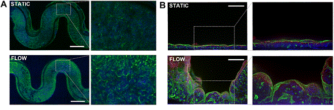
Figure 4. Immunofluorescence Staining of Caco-2 cell inside 3DP-µGut A) top view, nucleus in blue (DAPI and actin in green, bar = 750 μm B) cross-section view, nucleus in blue (DAPI), tight junctions (ZO-1) in red and actin in green, bar = 100 μm. From Delannoy et al. Lab Chip, 2025 [6].
Equipment for Gut-on-a-Chip Experiments
A gut-on-a-chip experiment requires a chip with the appropriate geometry, a flow source able to deliver microlitre-per-minute rates without pulsation, and a means of running that flow for days inside an incubator. Instrument selection follows the shear stress target and the intended culture duration.
Pressure-driven control with an in-line flow sensor sets flow rate in closed loop, which is the configuration used in the application notes above. The Flow EZ covers this function as a standalone unit.
The Omi automated organ-on-a-chip platform integrates flow control and recirculation in one enclosure sized to sit under a microscope or inside an incubator, for protocols running over several days.
Pre-configured cell perfusion and mechanical stimulation packages group the controller, sensors, reservoirs and tubing needed for perfusion and for cyclic stimulation respectively.
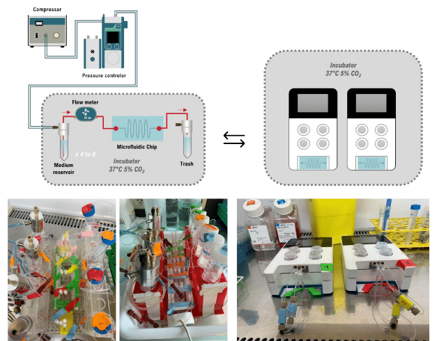
Figure 4. Flow EZ Perfusion Set-Up and Omi Platform with 3DP-µGut. From Delannoy et al. Lab Chip, 2025 [6].


Frequently Asked Questions
What is a gut-on-a-chip used for?
A gut-on-a-chip is used to study oral drug absorption and permeability, intestinal inflammation and inflammatory bowel disease, host–microbiome interactions, pathogen infection and nutrient transport. It uses human cells under flow rather than animal models or static wells.
How is peristalsis reproduced on a chip?
Peristalsis is reproduced by cyclic mechanical deformation of the membrane carrying the epithelium, typically through vacuum applied to side chambers. In the original human gut-on-a-chip this was 10 % strain at 0.15 Hz, applied together with luminal flow [9].
How does shear stress affect intestinal cells?
Low shear stress, on the order of 0.002 Pa, promotes Caco-2 polarisation, villus-like morphogenesis, tight junction assembly and mucus secretion. The response includes cytoskeletal remodelling and differentiation-associated gene expression [6], so shear is a cue and not merely medium replenishment.
Can the microbiome be co-cultured on a gut-on-a-chip?
Yes. A transluminal oxygen gradient in a gas-impermeable chip supports extended co-culture of aerobic and anaerobic human gut microbiota with living epithelium. A published anaerobic intestine-on-a-chip sustained over 200 operational taxonomic units from 11 genera [10].
Related Expertises
-
Expert Reviews: Basics of Microfluidics Liver-on-a-Chip: Models, Drug Metabolism and Applications Read more
-
Expert Reviews: Basics of Microfluidics Organ-on-a-Chip vs Organoids: Differences, Strengths and When to Combine Them Read more
-
Expert Reviews: Basics of Microfluidics Organ-on-Chip in Drug Development: A Human-Relevant Alternative to Animal Testing Read more
-
Expert Reviews: Basics of Microfluidics 5 Key Tips for Starting Organ-on-Chip Models Read more
-
Microfluidics Case Studies Gut-on-Chip Modeling: From Chip Development to Perfusion Read more
-
Microfluidic Application Notes Gut-on-Chip Model Development Using OOAC Platform, Omi Read more
-
Expert Reviews: Basics of Microfluidics Fluid Shear Stress in Cell Biology: Why and How to Control It Read more
-
Microfluidics White Papers A review of Organ on Chip Technology – A White Paper Read more
-
Microfluidic Application Notes Development of a human gut-on-chip to assess the effect of shear stress on intestinal functions Read more
References
- Bein A, Shin W, Jalili-Firoozinezhad S, Park MH, Sontheimer-Phelps A, Tovaglieri A, Chalkiadaki A, Kim HJ, Ingber DE. Microfluidic organ-on-a-chip models of human intestine. Cell Mol Gastroenterol Hepatol. 2018;5(4):659–668. doi:10.1016/j.jcmgh.2017.12.010
- Kasendra M, Tovaglieri A, Sontheimer-Phelps A, Jalili-Firoozinezhad S, Bein A, Chalkiadaki A, Scholl W, Zhang C, Rickner H, Richmond CA, Li H, Breault DT, Ingber DE. Development of a primary human Small Intestine-on-a-Chip using biopsy-derived organoids. Sci Rep. 2018 Feb 13;8(1):2871. doi: 10.1038/s41598-018-21201-7.
- Sambuy Y, De Angelis I, Ranaldi G, Scarino ML, Stammati A, Zucco F. The Caco-2 cell line as a model of the intestinal barrier: influence of cell and culture-related factors on Caco-2 cell functional characteristics. Cell Biol Toxicol. 2005;21(1):1–26. doi:10.1007/s10565-005-0085-6
- Toepke MW, Beebe DJ. PDMS absorption of small molecules and consequences in microfluidic applications. Lab Chip. 2006;6(12):1484–1486. doi:10.1039/b612140c
- Srinivasan B, Kolli AR, Esch MB, Abaci HE, Shuler ML, Hickman JJ. TEER measurement techniques for in vitro barrier model systems. J Lab Autom. 2015;20(2):107–126. doi:10.1177/2211068214561025
- Delannoy E, Burette A, Janel S, Poiret S, Deboosere N, Daniel C, A. Grassart. Gut-on-chip methodology based on 3D-printed molds: a cost-effective and accessible approach. Lab Chip. 2025; 25 (17): 4396–4409. doi:10.1039/D5LC00147A
- Kim HJ, Ingber DE. Gut-on-a-Chip microenvironment induces human intestinal cells to undergo villus differentiation. Integr Biol. 2013;5(9):1130–1140. doi:10.1039/c3ib40126j
- Sontheimer-Phelps A, Chou DB, Tovaglieri A, Ferrante TC, Duckworth T, Fadel C, Frismantas V, Sutherland AD, Jalili-Firoozinezhad S, Kasendra M, Stas E, Weaver JC, Richmond CA, Levy O, Prantil-Baun R, Breault DT, Ingber DE. Human colon-on-a-chip enables continuous in vitro analysis of colon mucus layer accumulation and physiology. Cell Mol Gastroenterol Hepatol. 2020;9(3):507–526. doi:10.1016/j.jcmgh.2019.11.008
- Kim HJ, Huh D, Hamilton G, Ingber DE. Human gut-on-a-chip inhabited by microbial flora that experiences intestinal peristalsis-like motions and flow. Lab Chip. 2012;12(12):2165–2174. doi:10.1039/c2lc40074j
- Jalili-Firoozinezhad S, Gazzaniga FS, Calamari EL, Camacho DM, Fadel CW, Bein A, Swenor B, Nestor B, Cronce MJ, Tovaglieri A, Levy O, Gregory KE, Breault DT, Cabral JMS, Kasper DL, Novak R, Ingber DE. A complex human gut microbiome cultured in an anaerobic intestine-on-a-chip. Nat Biomed Eng. 2019;3(7):520–531. doi:10.1038/s41551-019-0397-0
- Shin W, Wu A, Massidda MW, Foster C, Thomas N, Lee DW, Koh H, Ju Y, Kim J, Kim HJ. A robust longitudinal co-culture of obligate anaerobic gut microbiome with human intestinal epithelium in an anoxic-oxic interface-on-a-chip. Front Bioeng Biotechnol. 2019;7:13. doi:10.3389/fbioe.2019.00013
- Kim HJ, Li H, Collins JJ, Ingber DE. Contributions of microbiome and mechanical deformation to intestinal bacterial overgrowth and inflammation in a human gut-on-a-chip. Proc Natl Acad Sci USA. 2016;113(1):E7–E15. doi:10.1073/pnas.1522193112
- Nguyen TLA, Vieira-Silva S, Liston A, Raes J. How informative is the mouse for human gut microbiota research? Dis Model Mech. 2015;8(1):1–16. doi:10.1242/dmm.017400
- Ashammakhi N, Nasiri R, Barros NR, Tebon P, Thakor J, Goudie M, Shamloo A, Martin MG, Khademhosseini A. Gut-on-a-chip: Current progress and future opportunities. Biomaterials. 2020 Oct;255:120196. doi: 10.1016/j.biomaterials.2020.120196.
- Moerkens R, Mooiweer J, Smits E, Berg M, Ramírez-Sánchez AD, Modderman R, Puschhof J, Pleguezuelos-Manzano C, Barrett RJ, Wijmenga C, Jonkers IH, Withoff S. Gene expression profiling reveals enhanced nutrient and drug metabolism and maturation of hiPSC-derived intestine-on-chip relative to organoids and Transwells. Stem Cell Reports. 2025;20(12):102715. doi:10.1016/j.stemcr.2025.102715
- Shin W, Kim HJ. Intestinal barrier dysfunction orchestrates the onset of inflammatory host–microbiome cross-talk in a human gut inflammation-on-a-chip. Proc Natl Acad Sci USA. 2018;115(45):E10539–E10547. doi:10.1073/pnas.1810819115


