Mimicking in-vivo environments: biochemical and biomechanical stimulation
Cells are constantly exposed to biochemical stimulation from the early embryonic stage to adult life. The spatiotemporal regulation of these signals is essential as it determines cell fate, phenotype, metabolic activity as well as pathological behaviors. The fast response and high stability of Fluigent instruments make them the best solution available on the market to reproduce these complex variations in vitro.
Picture: Stable biochemical gradient
Advantages of creating an in-vivo environment for cell biology
Biochemical and biomechanical stimulation are two key types of signals that cells constantly receive from their environment, from the early embryonic stage to adult life. They play important roles in regulating cell behavior, phenotypes, metabolic activity and function.
Mimicking in-vivo environments by reproducing all the relevant chemical processes and biomechanical constraints in vitro is essential to inducing the right phenotype in cells, finalizing their maturation and maintaining homeostasis. The fast response and high stability of Fluigent instruments make them the best solution available on the market to reproduce these complex variations in vitro.
Benefits of using Fluigent’s instruments in creating a physiological microenvironment
Biochemical stimulation to mimic in-vivo environments
Temporal resolution: switches solutions rapidly (down to 20ms)
Spatial resolution: multiple flow focusing delivers gradients with one-cell diameter precision (cf. ref Benedetto et al)
Stable in time: pulse-free flow for up to 1 month
Programmable injection sequences: up to 10 distinct solutions
Sensitive and versatile: covers a large flow-rate range from nl/min to mL/min with a resolution of 1% of the measured value.
High throughput: perfuse up to 8 chips in parallel with specific medium and flow rate.
Migrating cells in dynamic chemical gradients generated by a microfluidic chamber (cells in gray, chemoattractant with fluorescein in green). Video courtesy of Prof. Satoshi Sawai (University of Tokyo, Japan)
Biomechanical stimulation to mimic in-vivo environments
Fast response: pulsatile flow (artery), steady laminar flow (veins, microvessels), quick and sharp mechanical stimulation
Stable over a large range of flow rates: covers a large flow-rate range from nl/min to mL/min with a resolution of 1% of the measured value.
Programmable pressure/flow rate profiles: cyclic mechanical strain (breathing, arterial blood pressure…) or complex pattern of mechanical stimulations
Sensitive and versatile: suitable for studying mechanical responses from cytoskeleton (pN) to tissue scale (arteries (P = 100mbar)).
High resolution: small pressure increments accessible by the system at any pressure (0.1 mbar – 7bar).
What are the possible applications?
Mimicking an in-vivo environment has many applications across a broad range of scientific fields, providing a useful tool for research and development in various domains.
Biochemical stimulation
Chemotaxis: study cell response to steady or dynamic chemical gradients.
Spatial control of the biochemical environment: co-streaming or successive injection of different fluids at a specific location.
Molecule characterization: identify a compound’s effect on biological processes from molecular (cytoskeleton) to tissue scale.
Drug screening: automate the injection time and sequence of compounds at given concentrations as well as periodic sampling at the system output.
Hyperoxia/hypoxia: control oxygen conditions.
Automated protocols: cell seeding, sequential injection, medium recirculation, immunostaining.
Surface coating
Immediate switching from actin to cofilin solution to study the depolymerization of actin filaments. (actin in red, cofilin in green). Video courtesy of Dr. Hugo Wioland (Institut Jacques Monod, France)
Biomechanical stimulation

Another way to imitate the in-vivo environment is through biomechanical stimulation:
Biomechanical/biophysical characterization of cytoskeleton, nucleus, cells, tissues, organs.
Morphogenesis: identify the biomechanical stimuli inducing cell proliferation, maturation and differentiation.
Mechanotaxis: study cell response to temporal or steady shear stress gradients.
Friction in biomedical devices: investigate frictional interactions between cells and the surface of the device to reduce inflammatory reactions.
Diagnosis based on biomechanical criteria: cell sorting/capture based on their deformability (e.g. isolate circulating tumor cells from blood sample), biomechanical characterization of patient samples (e.g. one symptom of falciparum malaria is the reduction in the deformability of infected red blood cells).
Hemodynamics, lymph dynamics: examine hydrodynamic properties of biological fluids in physiological conditions mimicking the in-vivo environment (relevant geometries imposed by the device design coupled to a consistent flow profile delivered by the flow controller)
Transport: quantify the transport of gas and nutrients under realistic conditions (flow rates, % of cell stretching (alveolus))
Related applications
- Expert Reviews: Basics of Microfluidics
Micropipette aspiration of cells and tissues
Read moreMicropipette aspiration is a powerful non-invasive technique to evaluate how biomechanical properties of single cells or tissue govern cell shape, cell response to mechanic stimuli, transition from nontumorigenic to tumorigenic state or morphogenesis
- Expert Reviews: Basics of Microfluidics
High Throughput Single Cell Analysis
Read moreMicrofluidics and lab-on-a-chip technology have emerged as the most promising approaches to address these challenges. These innovative technologies hold the potential to unlock new insights into single cell properties and their roles within populations.
Selected publication from our customers
- Wioland H et al, ADF/Cofilin accelerates actin dynamics by severing filaments and promoting their depolymerization at both ends. 2017. Curr Biol; 27(13):1956-1967.e7
- Tong et al, Crossed flow microfluidics for high throughput screening of bioactive chemical-cell interactions. 2017. Lab Chip;17(3):501-510.
- Nakajima A and Sawai S, Dissecting spatial and temporal sensing in Dictyostelium chemotaxis using a wave gradient generator. 2016.Methods Mol Biol; 1407:107-22.
- Nakajima A et al, The microfluidic lighthouse: an omnidirectional gradient generator. 2016. Lab Chip; 16(22):4382-4394
- Perez-Toralla K et al, FISH in chips: turning microfluidic fluorescence in situ hybridization into a quantitative and clinically reliable molecular diagnosis tool. 2015. Lab Chip; 15(3):811-22.
- Autebert J et al, High purity microfluidic sorting and analysis of circulating tumor cells: towards routine mutation detection. 2015. Lab Chip;15(9):2090-101.
- Schulze A et al, Dynamical feature extraction at the sensory periphery guides chemotaxis. 2015.Elife;4
- Benedetto A et al, Spatiotemporal control of gene expression using microfluidics. 2014. Lab Chip; 14(7):1336-47.
- Nakajima A et al, Rectified directional sensing in long-range cell migration. 2014. Nat Commun; 5:5367.
- Wilson K et al, Mechanisms of leading edge protrusion in interstitial migration. 2013.Nat Commun;4:2896.
- Startsev MA et al, Nanochannel pH gradient electrofocusing of proteins. 2013, Anal Chem.;85(15):7133-8
- Pernier J et al, Dimeric WH2 domains in Vibrio VopF promote actin filamenet barbed-end uncapping and assisted elongation. 2013, Nat Struct Mol Biol; 20(9): 1069-76
- Niman CS et al, Controlled microfluidic switching in arbitrary time-sequences with low drag. 2013, Lab Chip; 13(12):2389-96
- Niedermayer T et al, Intermittent deplolymerization of actin filaments is caused by photo-induced dimerization of actin protomers. 2012, Proc Natl Acad Sci USA;109(27):10769-74
- Jégou A et al, Individual actin filaments in a microfluidic flow reveal the mechanism of ATP hydrolysis and give insight into the properties of profilin. 2011, PLoS Biol;9(9): e1001161
- Saliba AE et al, Microfluidic sorting and multimodal typing of cancer cells in self-assembled magnetic arrays. 2010, Proc Natl Acad Sci USA; 107 (33):14524-9
- Bazargan V and Stoeber B, Moving temporary wall in microfluidic devices. 2008, Phys Rev Stat Nonlin Soft Matter Phys; 78(6 Pt 2): 066303
Related products
-
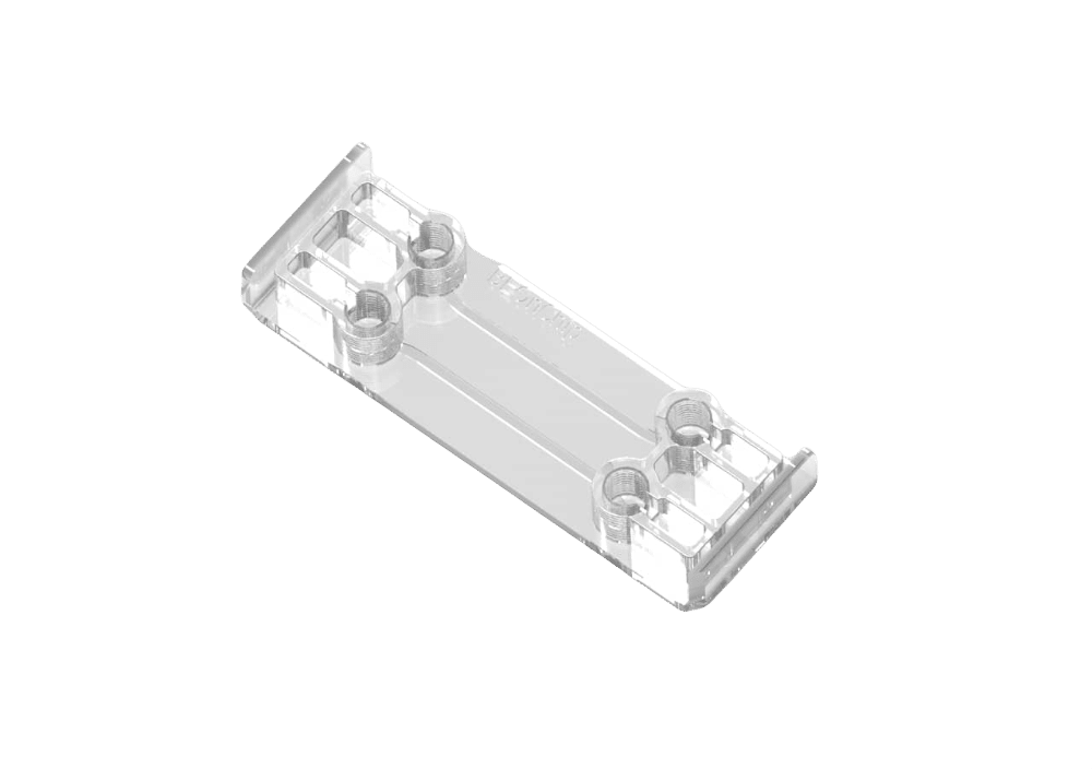
 Microfluidic cell culture chip Read
 -
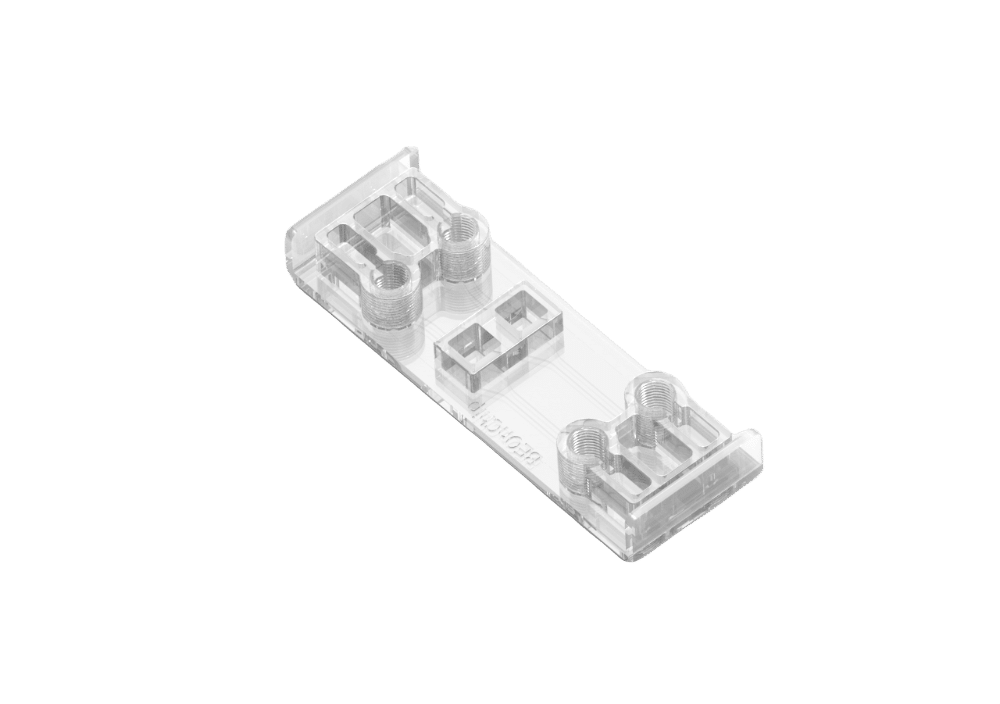
Air Liquid Interface Cell culture versatile chip Read
-
Flow gradient chip for 3D cell cultures Read
-
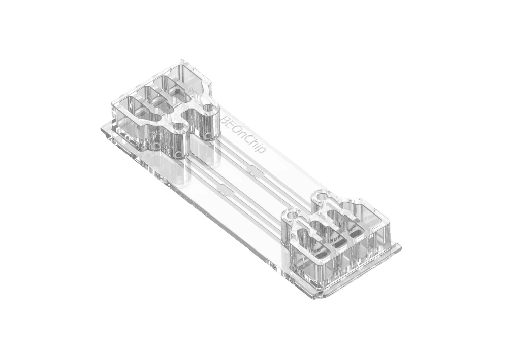
 Live Cell Imaging Chamber Read
 -
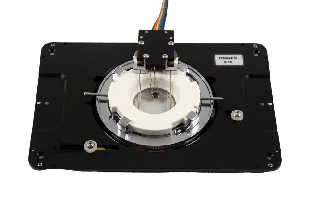
 Automated Multiplexed Imaging Platform Read
 -
Organ on Chip Perfusion Pack Read
-
High Throughput Cell Perfusion Pack Read
-
 Omi, automated organ-on-chip platform Read