• 简体中文
  • 한국어
  • Deutsch
Fluigent
  • Research
      • Flow EZ™ microfluidic flow controller
      • Microfluidic Research Equipment
      • Microfluidic Instruments
        • Omi, an Automated Organ-On-A-Chip Platform
        • Automated Perfusion System for Spatial Omics 
        • Microfluidic Pressure Based Flow Controller
        • Microfluidic Sensors
        • Microfluidic Valves
        • Pressure Control Reservoirs
        • Microfluidic Pressure Sources
        • Microfluidic Chips
        • Microfluidic Accessories
      • Software Solutions
        • Lab Integration Software
        • Real-Time Control & Lab Automation Software 
      • Microfluidic Packs
        • UV-crosslinked microcapsule production platform 
        • Encapsulation Platform for FACS
        • Microfluidic Complex Emulsion Production Platform
        • Microfluidic Application Packs
        • Microfluidic Starter Packs
      • Research Applications
        • Microfluidics for Cell Analysis
        • Microfluidics for Organ-on-chip Cell culture
        • Microfluidics for Droplet Generation
        • Microfluidics for Cell Biology
      • Subscribe to the newsletter
  • Industrial
      • Microfluidic OEM
      • Microfluidic OEM Devices
        • Microfluidic OEM Components
        • Customizable OEM Flow Control Modules
      • Fully Custom Microfluidic Device
      • Custom microfluidic device
      • Technologies
        • A Microfluidic Pressure Controller Comparison for Your Ultimate Fluid Control System
        • 5 reasons to choose OEM pressure controllers over OEM syringe pumps for microfluidic applications
        • Microfluidic recirculation system 
        • DFC, “Self-Learning” Microfluidic Flow Control Algorithm
        • Non-Intrusive Flow Sensing Technology
        • Compact All-In-One Microfluidic Micropump
        • Liquid Stirring Solutions
      • Industrial Applications
        • Combining Microfluidics and Spectroscopy
        • Valve Automation with the F-OEM for Microfluidic Applications
        • Localization microscopy and flow control for multiplexing 
        • Contamination-free Liquid Handling System
        • Microfluidic Drug Discovery 
        • Flow Expertise for Cell Encapsulation and Single-Cell Analysis
        • Droplet Digital PCR (ddPCR)
      • Subscribe to the newsletter
  • Markets & Applications
    • Microfluidics in Life Science
    • Microfluidics for Pharmaceutical Applications
    • Microfluidics for Food testing & Agriculture
    • Microfluidics in Cosmetics
    • Microfluidics in Water analysis
  • Company
    • About us
    • Fluigent’s Academic Partners
      • Scientific Partners
      • Fluigent’s Brand Ambassadors
      • Center Partners
    • Team
    • News
    • Events & Webinars
    • Fluigent Newsletter
    • Fluigent’s Distributors
    • Careers
  • Resources & Support
      • Expert Reviews: Basics of Microfluidics
        • General overview of microfluidics
        • Advantages of pressure-based microfluidics
        • Microfluidics tips
        • Droplet & Particle Generation
        • Microfluidic cell biology
        • Industrial / OEM Expertise
        • Funded research program
      • FAQ
      • Videos
        • Expertise videos
        • Product presentation videos
        • Tutorial videos
      • Expertise
        • Videos
        • Microfluidics Article Reviews
        • Microfluidic Application Notes
        • Microfluidics case studies
        • Interviews & Testimonials
        • Microfluidics White Papers
      • Documentation
        • CAD
        • Fluigent Catalog
        • Fluigent products manual
        • Fluigent Products Datasheets
        • Safety datasheet
        • Fluigent Media Kit product icons & images
      • Microfluidic Calculators
        • Shear Stress Calculator
        • Pressure & Flow Rate Calculator
        • Droplet Size Calculator
      • Download software
        • FEZ and Link Firmware Updater​
        • OxyGEN
        • Software Development Kit
        • Discontinued software
      • Subscribe to the newsletter
  • Webshop
  • Contact us
Home » Resources » Expertise » Microfluidic Application Notes » Cartilage-on-a-chip, an example of complex mechanical stimulation using Fluigent’s technology

Cartilage-on-a-chip, an example of complex mechanical stimulation using Fluigent’s technology    

In understanding the processes responsible for osteoarthritis or other cartilage diseases, exploring the response of chondrocytes to external mechanical and chemical stimulations can lead to innovative therapies. While traditional approaches have allowed the scientific community to highlight some particularly interesting behaviors of chondrocytes, their lack of physiological relevance can lead to false conclusions.

In this application note, we report a new method for 3D chondrocyte culture using a microfluidic platform associated with  Fluigent products, enabling researchers to create a relevant cartilage model subject to complex mechanical stimulation.

This application note was made possible thanks to Séverine Le Gac and her team at Applied Microfluidics for BioEngineering Research (AMBER). For more information, visit the team website.

Download the application note

What’s the value of simulating complex mechanical stimulation?

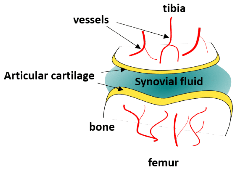
Articular cartilage is a type of connective tissue located at the end of long bones to protect them from friction and to help them support heavy loads (Fig 1). It is usually bathed in synovial fluid, and is therefore subject to mechanical and biochemical activity. 

Biochemical or mechanical breakdown of the cartilage results in a disease called osteoarthritis, the most common joint disease, which affects millions of people worldwide. Numerous models have been developed by scientists to understand the processes underlying this disorder, with the goal of finding an optimized cure. However, current methods usually rely on 2D cultures of chondrocyte cells in static conditions, which is physiologically far from the in-vivo 3D assembly of the cells inside an extracellular matrix constantly exposed to fluid flows. 

schema-compressive-stress-cell
Figure 1: Schematic of the knee joint.  
mimic compression
Figure 2: PDMS membrane deformation by application of pressure in the actuation chambers 

Microfluidics is of growing interest in the scientific community, as it allows for perfused 3D culture of cells, enabling the development of highly physiologically relevant models in systems referred to as organ-on-a-chip. In these models, complex mechanical stimulations can be reproduced by the controlled injection of fluids into the system. 

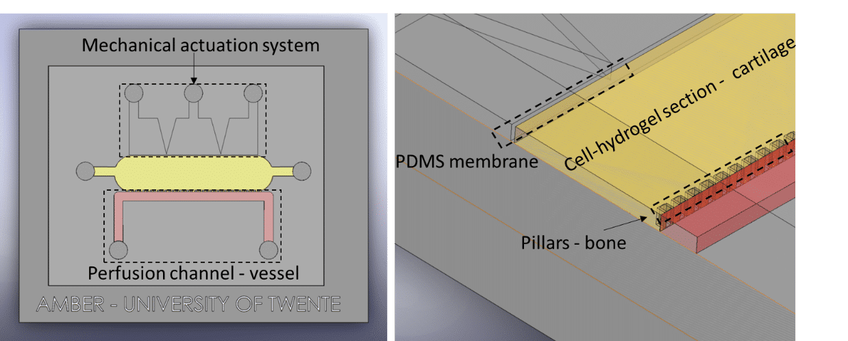
Séverine Le Gac’s team developed a cartilage model in which an agarose hydrogel containing cultured human chondrocytes is subject to compression through the pressurization of actuation chambers to trigger membrane deformation (Fig 2). The effect of this advanced mechanical stimulation on cells was observed. 

Generating complex mechanical stimulation

Fluigent instruments

microfluidic flow control system

Microfluidic Flow Control System

Read more

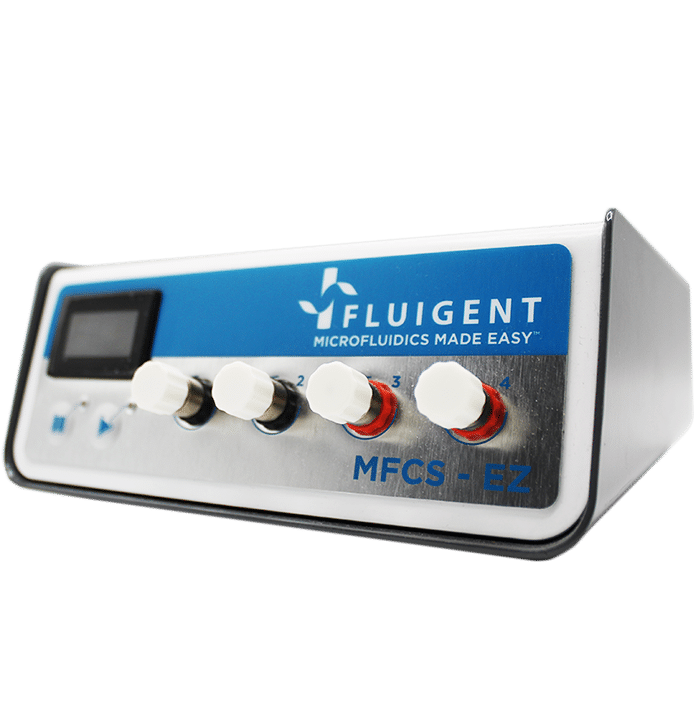
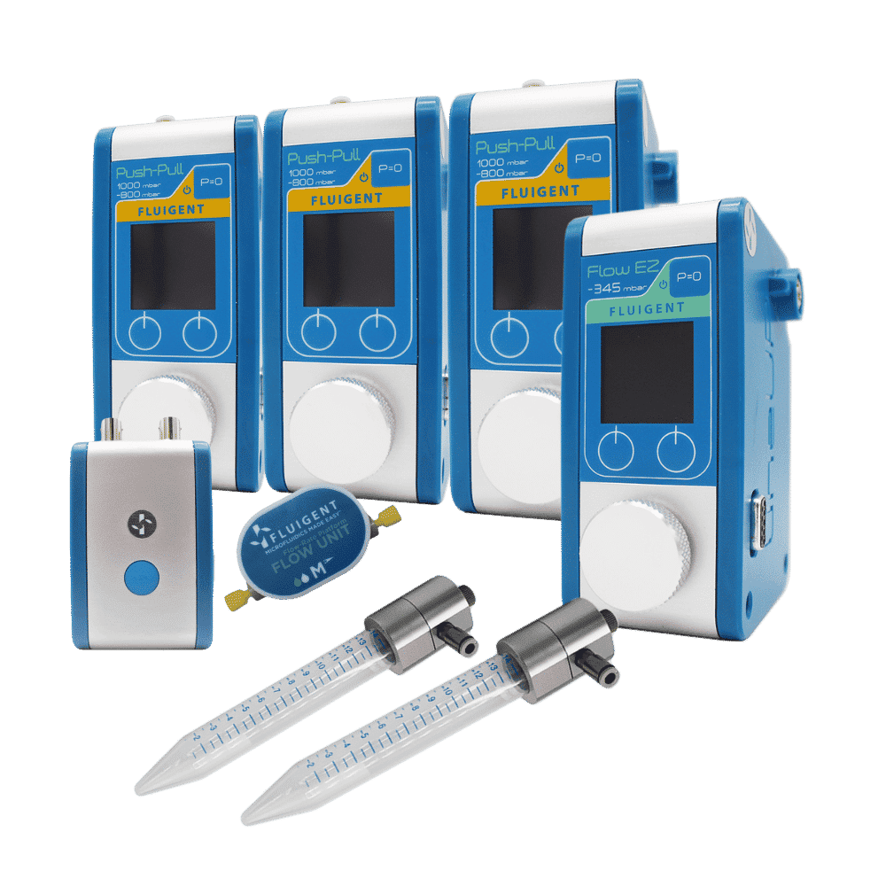
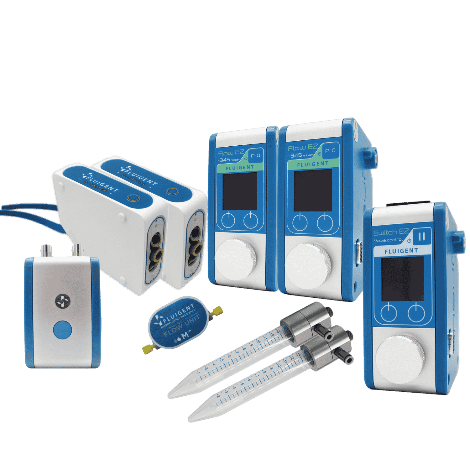
Pressure generator: An MFCS-EZ pressure controller was used for fluid injection with an excellent response time and high stability. Pressure ranging from 0 to 1500 mbar was generated. 

MICROFLUIDIC SAMPLING VALVE 2 switch

Microfluidic Sampling Valve

Read more

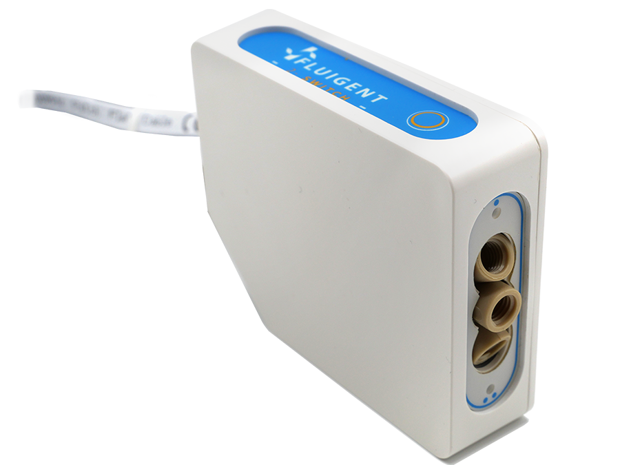
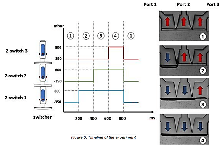
Valves: Three 2-switch valves were connected to the pressure generator and a vacuum pump to apply  positive or negative pressure to the three actuation chambers and generate complex mechanical stimulation. The valves were connected to a switchboard to allow monitoring on the computer. 

Real-Time Control & Lab Automation Software 

Read more

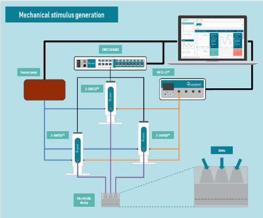
Software: Our automation software was used to predefine and automate the pressure levels, enabling the production of complex patterns inside the chip. 

The Fluigent setup is shown in Fig 3. 

Fig 3: Fluigent instrument setup 

Other instruments

Microfluidic chip: Conventional soft-lithography techniques were used to fabricate the chip, made of poly-dimethyl-siloxane (PDMS) and bonded to a PDMS-coated glass slide. This device allows for 3D culture of chondrocytes under dynamic conditions (nutrient supply) and enables the application of a complex mechanical stimulation by deforming the PDMS membrane using pressure (Fig 4). 

Camera: An ORCA-flash 4.0 LT from Hamamatsu Photonics was used to visualize the effect of pressure on the PDMS membrane. 

Compressive stress
Fig 4: Schematic of the microfluidic chip 

Methods to create complex mechanical stimulation

Experiments were done to verify the cellular viability of chondrocytes upon application of advanced mechanical stimulation. First, a culture of chondrocyte cells isolated from a patient undergoing knee replacement was done. Then cells were cultured inside the microfluidic chamber, with or without (control) mechanical stimuli via application of pressure in the actuation chambers. Several pressures were tested. Finally, a live/dead assay was performed using fluorescent probes to assess cell viability. 

complex mechanical stimulation - pressure actuation
Fig 5: Experimental timeline of pressure actuation 

Partial results

It was demonstrated that cell viability was not affected by the membrane deformation. The ratio of living cells to dead cells was similar in the case of chondrocytes subject to complex mechanical stimulation and in the control test. 

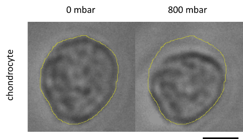
In addition, a study was performed of the cell deformation due to application of pressure in the actuation chambers, and showed that it was not uniform in the matrix, with a maximum deformation value of 13% close to the membrane, decreasing along the matrix’s depth (Fig 6). 

Chondrocyte deformation
Figure 6: Chondrocyte deformation: a) no pressure; b) 800 mbar of compression. (Scale: 7µm)

Conclusion

Fluigent products were used as an essential tool to develop a physiologically relevant 3D cartilage model and create a complex mechanical stimulation. The application of pressure in the actuation chambers was demonstrated to have an effect on cell shape, but no effect on cell viability was found. More analysis will be done to evaluate the response of chondrocytes to this deformation. 

omi organ models development

Related resources

  • A guide to Organs-on-chips (OoC) technology

    White paper: Organ on Chip

    Read more
  • Microfluidics White Papers

    Microfluidic white paper – A review of Organ on Chip Technology 

    Read more
  • Microfluidics White Papers

    An exploration of Microfluidic technology and fluid handling 

    Read more

Related products

  • Mechanical Stimulation Pack Read
  • pack recirculation products
    Microfluidic Recirculation Pack Read
  • CELL PERFUSION PACK
    High Throughput Cell Perfusion Pack Read
  • microfluidic flow control system
    Microfluidic Flow Control System Read

For more information or a technical discussion

Contact us
Logo fluigent green and blue

67 avenue de Fontainebleau
94 270 Le Kremlin-Bicêtre

Research

  • Microfluidic Research Applications
  • Instruments
  • Software solutions
  • Packages

Industrial

  • Products

Resources

  • Microfluidic Application Notes
  • Microfluidics case studies
  • Expert Reviews: Basics of Microfluidics
  • Interviews & Testimonials

Support & Tools

  • Documentation
  • Download software

Company

  • About us
  • Team
  • Events & Webinars
  • Newsletter
  • Fluigent’s Distributors
  • Careers

Legal

  • Terms & Conditions of Sale
  • Legal Terms & Privacy Policy