A Microfluidic approach for modeling the blood-brain tumor barrier
The microvasculature of the central nervous system (CNS) is essential for maintaining the integrity of the brain. The blood-brain barrier (BBB) is a selective, semi-permeable barrier that controls the passage of molecules from the blood to the brain. Because of its restrictive properties, the BBB constitutes a major obstacle to effective drug delivery to the CNS [1, 2]. This barrier comprises several cell types: endothelial cells, pericytes, and glial cells, including astrocytes and microglia. The close collaboration of the cells is fundamental for the proper functioning of the BBB. The presence of tumor cells contributes to disrupting the BBB and giving rise to the blood-brain tumor barrier (BBTB), a heterogeneous vasculature characterized by uneven permeability and active efflux of molecules [3]. Consequently, substantial research has focused on developing models that approach the complexity of the BBTB to study strategies to facilitate the transport of therapeutics in this microenvironment.
To overcome these challenges, N. Barin et al. introduced a microfluidic 3D printed in vitro model that integrates vessel architecture with micrometric resolution, supports multicellular culture, and is compatible with in-chip fabrication. This approach involves two-photon polymerization (2PP) to create a pertinent microvessel platform.
Enabled by Fluigent’s pressure-based flow control, it ensures precise, stable and reproducible conditions under dynamic flow conditions.
Key Takeaways
- The blood-brain tumor barrier (BBTB) forms when tumor cells disrupt the BBB, creating a leaky, heterogeneous vasculature that limits effective drug delivery to brain tumors.
- Researchers at Delft University of Technology and the ErasmusMC Cancer Institute used two-photon polymerization (2PP) to fabricate microporous capillary scaffolds (µPCs) with vessel-scale dimensions (40-50 µm).
- The µPC platform supports co- and tri-culture of endothelial cells, pericytes, and glioma cells, closely reproducing the cellular complexity of the BBTB.
- Fluigent’s pressure-driven flow control (Flow EZ and Flow Unit sensors) delivered stable, physiologically relevant shear stress (6.5-7.8 dyn/cm²) to the model under dynamic culture conditions.
- The resulting in vitro model offers a reproducible, physiologically relevant tool for studying BBTB biology and testing drug candidates for brain tumors.
The blood-brain tumor barrier in brain cancer
Under normal conditions, the physiological characteristics of the BBB prevent the leakage of molecules due to the presence of tight junctions between the endothelial cells. The only molecules that can passively diffuse across the BBB are gases (ex: oxygen and carbon dioxide) and small lipid-soluble molecules with a low molecular weight (<400 Da) [4]. The transporters are located at the abluminal and luminal phases of the endothelial cells [5].
Brain tumors are a heterogeneous group of CNS neoplasms classified as either primary, originating within the brain, or secondary, resulting from the metastasis of peripheral tumors. Glioblastoma is the most common primary malignant brain tumor in adults, carrying a median overall survival of approximately 15 months despite aggressive therapy [6]. During tumor progression, the BBB is disrupted and presents aberrant angiogenesis, leading to a leaky barrier known as the blood-brain tumor barrier (BBTB) [7]. The BBTB is also characterized by aberrant pericyte distribution, which are perivascular cells essential for maintaining BBB integrity, and loss of astrocytic endfeet [8].
Primary brain tumors, including gliomas, disrupt the BBB’s integrity by altering normal vascular architecture [9]. Regarding metastatic brain tumors, the BBB is partially disrupted, with functional efflux transporters limiting drug delivery into the parenchyma. In this context, the successful treatment of the patient is challenging (Figure 1) [10].

Figure 1: Comparison of the neurovascular unit of the blood-brain barrier and blood-brain tumor barrier [11].
Modeling the blood-brain tumor barrier
The BBB is the main interface between the blood and the brain parenchyma. The main process of vascular network growth and remodeling takes place after the initiation of blood circulation. The BBB is exposed to mechanical forces derived from the blood flow, including shear stress, axial stress, and circumferential stress. These hemodynamic forces significantly affect the mechanics and morphology of brain blood vessels [12, 13]. In brain tumors, abnormal vascular architecture alters local flow patterns and contributes to the heterogeneous permeability characteristic of the BBTB [3]. It’s crucial to include these forces to recreate a pertinent BBTB model.
Nowadays, the in vitro modeling of the BBB and BBTB remains challenging because of its complexity. Existing preclinical models mainly consist of two-dimensional cell cultures and animal studies; however, both approaches fail to fully recapitulate human physiology, disease complexity or have limited throughput[14]. Microfluidic cell cultures have become essential to recreate accurate in vitro models of the BBTB. Microfluidic systems offer precise control of the fluid and allow to provide a dynamic microenvironment and the shear stress needed to mimic physiological conditions. Exposing endothelial cells to flow for the modeling of the BBB helps maintain cell morphology, function, and barrier properties [15]. Regarding the modeling of the BBTB, it is difficult to recreate the cell complexity of the barrier and the geometry of the vessels of the brain.
Why was a new blood-brain tumor barrier model needed?
Paper: N. Barin et al., “Two-Photon Polymerized Microvascular Environments for Multicellular Modeling of the Blood-Brain Tumor Barrier,” ADVANCED MATERIALS TECHNOLOGIES, vol. 11, no. 8, 2026-01-16 2026, doi: 10.1002/admt.202502614.
To overcome the limitations of the already available models, associate professor Angelo Accardo, PhD student Nastaran Barin and professor Pim French from Delft University of Technology and the ErasmusMC Cancer Institute (The Netherlands), set out to develop a more physiologically relevant BBTB model.
In their study published in Advanced Materials Technologies in 2026, and featured on its cover, the researchers employed two-photon polymerization (2PP) to fabricate ultrafine vascular scaffolds with precisely engineered pores, enabling both human umbilical vein endothelial cells (HUVECs) and human cerebral microvascular endothelial cells (hCMECs) to proliferate throughout the microporous capillary network. In addition to these endothelial cells, they successfully established a co- and tri-culture with pericytes and glioma cells [16].
Methodology: How to model a 3D blood-brain tumor barrier in dynamic conditions
The BBTB dynamic model developed in this study is based on 2PP to form a designed platform that includes a precise geometry for the vessel formation. The platform is based on a microporous tube-like capillary scaffold (µPC) (Figure 2), that guides the attachment and growth of the endothelial cells. The diameter chosen (40-50 µm) in this study is close to the dimensions of brain capillaries of the BBTB (15-30 µm). µPCs were fabricated directly inside a microfluidic chip by 2PP using a Nanoscribe Photonic Professional GT+ printer and IP-Visio, a methacrylate photosensitive polymer, which is biocompatible and low-autofluorescent. Different fabrication workflows were applied for single- and dual-channel chips, combining dip-in laser lithography (DiLL) and oil-immersion configurations to accommodate chip geometry and seal the structures.
To ensure precise and stable control of both fluidic and mechanical conditions, the system relies on pressure-driven flow provided by Fluigent flow control solutions. In this setup, the Flow EZ is used to generate a shear stress required to reproduce physiological conditions at the BBTB. Moreover, the setup included one Fluigent Flow Unit sensors for real-time monitoring of flow rates upstream of the chip.
To explore the potential for dynamic cell culture applications, 2PP-fabricated µPCs and support structures are fabricated directly into commercially available microfluidic chips. This integration is carried out using two different types of microfluidic chips (Figure 2A,B). The first type is a single-channel chip that allows direct flow inside the µPCs (Figure 2A), where endothelial cells and culture medium are introduced via the same channel. In the dual-channel chip configuration (Figure 2B), one channel provides access to the external surface of µPCs and is used to introduce endothelial cells.. The second channel enables internal fluid flow through the µPCs, exposing the inner surface of endothelial cells to media under physiological flow conditions. HUVECs or hCMECs were seeded in the structures previously coated with a solution of 5% collagen type I and cultured under static or dynamic conditions.
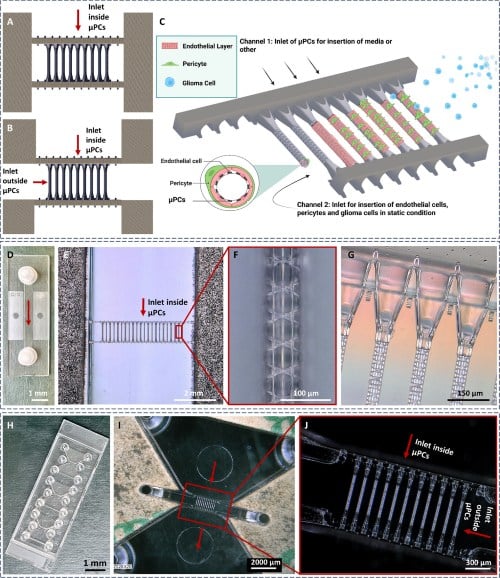
Figure 2: Design and fabrication of 3D µPCs. Schematics of the single-channel (A) and dual-channel (B–C) microfluidic chip designs, illustrating internal and external perfusion capabilities and the potential for multicellular culture. Images of the fabricated single-channel chip (D–G) and dual-channel chip (H–J), including optical views of the 2PP-fabricated µPCs, supporting walls, flow direction, and µPC inlets.
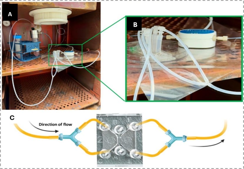
Figure 3: Microfluidic setup for dual-channel chips. A) Complete setup with chip, reservoir, waste container, flow sensor, and tubing placed inside the incubator. B) Zoomed-in view of the microfluidic chip connected to tubing for flow culture. C) Schematic illustration of inlet and outlet tubing connections used to maintain continuous flow.
Results: A pertinent development of in vitro BBTB using the µPC platform
The first step of this study was to characterize the cells on the µPCs in static conditions to study the development of the in vitro vessel-like structures and the impact of glioma cells on the model. The tri-culture system comprised endothelial cells (hCMEC/D3), pericytes, and glioma cells (U87) to form a pertinent and complex BBTB model. After 8 days of culture, the cellular organization within the system was visualized using immunofluorescence staining of cell-specific markers and confocal microscopy.
The presence of CD31-positive cells (Figure 4A) indicates the presence of endothelial cells lining the scaffold. This adhesion protein is involved in maintaining BBB integrity [17]. Moreover, positive staining for PDGFR-β, a marker expressed by mural cells, confirmed the successful adhesion and perivascular localization of pericytes around the endothelial cells within the µPCs while preserving the integrity of the vessel-like architecture. In the context of BBTB modeling, the incorporation of glioma cells was further validated by the detection of S100-positive cells, confirming their successful attachment to the model (Figure 4B). The quantitative analysis of the CD31 intensity between monoculture and co-culture conditions (Figure 4C) highlighted that the presence of glioma cells disrupted the endothelial junctions also in the presence of stabilizing pericytes, as reported in the literature [3, 18].

Figure 4: Endothelial barrier integrity on µPCs under static culture. Representative images of pericyte–endothelial co-culture (A) and U87 glioma–endothelial–pericyte tri-culture (B) on µPCs. (C) Quantification of CD31 fluorescence intensity in hCMEC/D3 cells from three independent experiments (n = 3).
Regarding the dynamic culture conditions, HUVEC cells were cultured using the Flow EZ pressure controller along with one Fluigent Flow Unit sensor (medium model) to apply a relevant flow. To be close to the brain capillaries shear stress (1–15 dyn/cm2 [19]), the µPCs models were submitted to the flow rates and resulting wall shear stress values summarized in Table 1, which remained within the physiological range. Under biomimetic flow conditions in a single-channel perfusion, endothelial cells uniformly colonized the surfaces of the scaffold (Figure 5B, C and D). Using the dual-channel chip, strong CD31 signaling was visible, indicating the formation of intercellular junctions and endothelial identity (Figure 5G). Importantly, the combination of the flow and the µPC platform in a chip allows the development of pertinent vessel-like structures with a physiological shear stress.
| Parameter | µPC model | Physiological reference |
|---|---|---|
| Vessel diameter | 40–50 µm | 15–30 µm (brain capillaries) |
| Flow rate – single-channel chip | 12 µL/min | _ |
| Flow rate – dual-channel chip | 5 µL/min | _ |
| Wall shear stress – single-channel chip | 7.8 dyn/cm² | 1–15 dyn/cm² (physiological range) |
| Wall shear stress – dual-channel chip | 6.5 dyn/cm² | 1–15 dyn/cm² (physiological range) |
Table 1: Flow parameters applied to the µPC model and their physiological reference values.

Figure 5: Endothelial cell culture on 2PP-fabricated µPCs under dynamic flow. Optical images of the single-channel (A) and dual-channel (E) microfluidic chips showing flow direction. Representative immunofluorescence images of HUVECs (B–G) and hCMEC/D3 cells (H) cultured on µPCs under flow, demonstrating endothelial coverage of the 3D µPC surfaces with CD31 or actin staining.
Conclusion: the µPC platform is an efficient tool for BBTB modeling
In this study, researchers from Delft University of Technology and the ErasmusMC Cancer Institute (The Netherlands) developed a pertinent model to study the BBTB. Compared to existing in vitro models, the µPC platform combined with the microfluidic system offers several distinct advantages. Unlike conventional microfluidic chip-based systems, it provides capillary-scale vessel dimensions and direct access to both the luminal and abluminal sides of the endothelium. The use of a pressure controller allows the exposure of the cells to a relevant shear stress, increasing the biomimetic properties of the system. µPC design enables highly controlled and reproducible vessel architecture with consistent flow dynamics. Overall, the platform in dynamic conditions would be an essential tool for introducing additional cell types to increase the complexity of the model, test new drug candidates for brain tumors, and evaluate their passage through the BBTB.
Frequently Asked Questions
What is the blood-brain tumor barrier (BBTB)?
The BBTB is the altered, leaky vasculature that forms when tumor cells disrupt the blood-brain barrier (BBB). It is characterized by uneven permeability, aberrant pericyte distribution, and loss of astrocytic endfeet, which together limit how well drugs reach brain tumors.
How did the researchers build the BBTB model?
The team used two-photon polymerization (2PP) to fabricate microporous capillary scaffolds (µPCs) with vessel-scale diameters (40–50 µm) directly inside microfluidic chips. Endothelial cells, pericytes, and glioma cells were then cultured on these scaffolds to reproduce the BBTB’s multicellular architecture.
What role did flow control play in the model?
Fluigent’s pressure-driven Flow EZ controller and Flow Unit sensors delivered stable, tunable flow, exposing the endothelial cells to wall shear stresses of 6.5–7.8 dyn/cm², within the 1–15 dyn/cm² range measured in real brain capillaries.
Why does this model matter for brain cancer research?
By combining capillary-scale geometry, multicellular co-culture, and physiological shear stress, the µPC platform gives researchers a more realistic tool to study BBTB biology and to test how well new drug candidates cross the barrier into brain tumors.
Related Solutions


Related Expertises
-
Expert Reviews: Basics of Microfluidics Cancer-on-Chip: Modeling the Tumor Microenvironment with Microfluidics Read more
-
Microfluidics Case Studies Biomechanics of Perfused Kidney-on-Chip Model: Effects of Shear Stress and Pressure Read more
-
Expert Reviews: Basics of Microfluidics Organ-on-Chip in Drug Development: A Human-Relevant Alternative to Animal Testing Read more
-
Expert Reviews: Basics of Microfluidics 5 Key Tips for Starting Organ-on-Chip Models Read more
-
Microfluidics Case Studies Mimicking tumor microenvironment using a 3D microfluidic model to improve cancer investigations Read more
-
Microfluidics Article Reviews Human Blood Brain Barrier (BBB) permeability -on-chip assessment Read more
-
Microfluidics White Papers A review of Organ on Chip Technology – A White Paper Read more
-
Expert Reviews: Basics of Microfluidics Why Control Shear Stress in Cell Biology? Read more
-
Microfluidic Application Notes Development of a human gut-on-chip to assess the effect of shear stress on intestinal functions Read more
-
Microfluidic Application Notes Peristaltic Pump vs Pressure-Based Microfluidic Flow Control for Organ on Chip applications Read more
-
Expert Reviews: Basics of Microfluidics Mimicking in-vivo environments: biochemical and biomechanical stimulation Read more
-
Expert Reviews: Basics of Microfluidics The Importance of Flow Control Stability in Microfluidics Read more
References
[1] R. Daneman and A. Prat, “The blood-brain barrier,” (in eng), Cold Spring Harbor Perspectives in Biology, vol. 7, no. 1, p. a020412, 2015/01/05/ 2015, doi: 10.1101/cshperspect.a020412.
[2] D. Wu, Q. Chen, X. Chen, F. Han, Z. Chen, and Y. Wang, “The blood-brain barrier: structure, regulation, and drug delivery,” SIGNAL TRANSDUCTION AND TARGETED THERAPY, vol. 8, no. 1, 2023-05-25 2023, Art no. 217, doi: 10.1038/s41392-023-01481-w.
[3] C. Arvanitis, G. Ferraro, and R. Jain, “The blood-brain barrier and blood-tumour barrier in brain tumours and metastases,” NATURE REVIEWS CANCER, vol. 20, no. 1, pp. 26-41, 2020-01-01 2020, doi: 10.1038/s41568-019-0205-x.
[4] W. A. Banks and C. L. Farrell, “Impaired transport of leptin across the blood-brain barrier in obesity is acquired and reversible,” American Journal of Physiology-Endocrinology and Metabolism, vol. 285, no. 1, 2003 Jul 01, doi: 10.1152/ajpendo.00468.2002.
[5] H. Kadry, B. Noorani, and L. Cucullo, “A blood–brain barrier overview on structure, function, impairment, and biomarkers of integrity,” Fluids and Barriers of the CNS, vol. 17, no. 1, p. 69, 2020/11/18/ 2020, doi: 10.1186/s12987-020-00230-3.
[6] J. Sung and K. Hwang, “Glioblastoma: epidemiology, molecular pathogenesis, diagnosis, management, and therapeutic resistance,” MOLECULAR BIOMEDICINE, vol. 7, no. 1, 2026-05-08 2026, Art no. 63, doi: 10.1186/s43556-026-00467-8.
[7] S. Liebner, R. M. Dijkhuizen, Y. Reiss, K. H. Plate, D. Agalliu, and G. Constantin, “Functional morphology of the blood-brain barrier in health and disease,” (in eng), Acta Neuropathologica, vol. 135, no. 3, pp. 311-336, 2018/03// 2018, doi: 10.1007/s00401-018-1815-1.
[8] L. Dubois et al., “Gliomas and the vascular fragility of the blood brain barrier,” FRONTIERS IN CELLULAR NEUROSCIENCE, vol. 8, 2014-12-12 2014, Art no. 418, doi: 10.3389/fncel.2014.00418.
[9] M. Ahmed, M. Canney, A. Carpentier, and A. Idbaih, “International meeting of the French society of neurology 2023 Overcoming the blood brain barrier in glioblastoma: Status and future perspective,” REVUE NEUROLOGIQUE, vol. 179, no. 5, pp. 430-436, 2023-05-15 2023, doi: 10.1016/j.neurol.2023.03.013.
[10] D. Masri et al., “The Role of Blood-Brain Barrier in Brain Cancer: From Pathophysiology to Therapeutic Approaches,” CANCER CONTROL, vol. 33, 2026-01-01 2026, Art no. 10732748261443870, doi: 10.1177/10732748261443870.
[11] B. Allen and C. Limoli, “Breaking barriers: Neurodegenerative repercussions of radiotherapy induced damage on the blood-brain and blood-tumor barrier,” FREE RADICAL BIOLOGY AND MEDICINE, vol. 178, pp. 189-201, 2021-12-09 2022, doi: 10.1016/j.freeradbiomed.2021.12.002.
[12] I. E. Hoefer, B. den Adel, and M. J. A. P. Daemen, “Biomechanical factors as triggers of vascular growth,” (in eng), Cardiovascular Research, vol. 99, no. 2, pp. 276-283, 2013/07/15/ 2013, doi: 10.1093/cvr/cvt089.
[13] P. Campinho, A. Vilfan, and J. Vermot, “Blood Flow Forces in Shaping the Vascular System: A Focus on Endothelial Cell Behavior,” Frontiers in Physiology, vol. 11, p. 552, 2020/06/05/ 2020, doi: 10.3389/fphys.2020.00552.
[14] D. Stanimirovic, M. Bani-Yaghoub, M. Perkins, and A. Haqqani, “Blood-brain barrier models: in vitro to in vivo translation in preclinical development of CNS-targeting biotherapeutics,” EXPERT OPINION ON DRUG DISCOVERY, vol. 10, no. 2, pp. 141-155, 2015-02-01 2015, doi: 10.1517/17460441.2015.974545.
[15] L. Cucullo, M. Hossain, V. Puvenna, N. Marchi, and D. Janigro, “The role of shear stress in Blood-Brain Barrier endothelial physiology,” BMC NEUROSCIENCE, vol. 12, 2011-05-11 2011, Art no. 40, doi: 10.1186/1471-2202-12-40.
[16] N. Barin et al., “Two-Photon Polymerized Microvascular Environments for Multicellular Modeling of the Blood-Brain Tumor Barrier,” ADVANCED MATERIALS TECHNOLOGIES, vol. 11, no. 8, 2026-01-16 2026, doi: 10.1002/admt.202502614.
[17] I. Wimmer et al., “PECAM-1 Stabilizes Blood-Brain Barrier Integrity and Favors Paracellular T-Cell Diapedesis Across the Blood-Brain Barrier During Neuroinflammation,” (in en), Frontiers in Immunology, vol. 10, 2019 2019, doi: 10.3389/fimmu.2019.00711.
[18] F. Mo, A. Pellerino, R. Soffietti, and R. Ruda, “Blood-Brain Barrier in Brain Tumors: Biology and Clinical Relevance,” INTERNATIONAL JOURNAL OF MOLECULAR SCIENCES, vol. 22, no. 23, 2021-12-01 2021, Art no. 12654, doi: 10.3390/ijms222312654.
[19] X. Wang et al., “Advances on fluid shear stress regulating blood-brain barrier,” MICROVASCULAR RESEARCH, vol. 128, 2020-03-01 2020, Art no. 103930, doi: 10.1016/j.mvr.2019.103930.