Cancer Cell Analysis Made Easy with Aria: cell Capture and Labeling
The invasiveness of cancer cells is a crucial indicator of a primary tumor's metastasizing capability. Detecting and quantifying cancer cell invasion is vital for predicting a patient's survival rate and evaluating the impact of anti-cancer compounds on cancer progression.
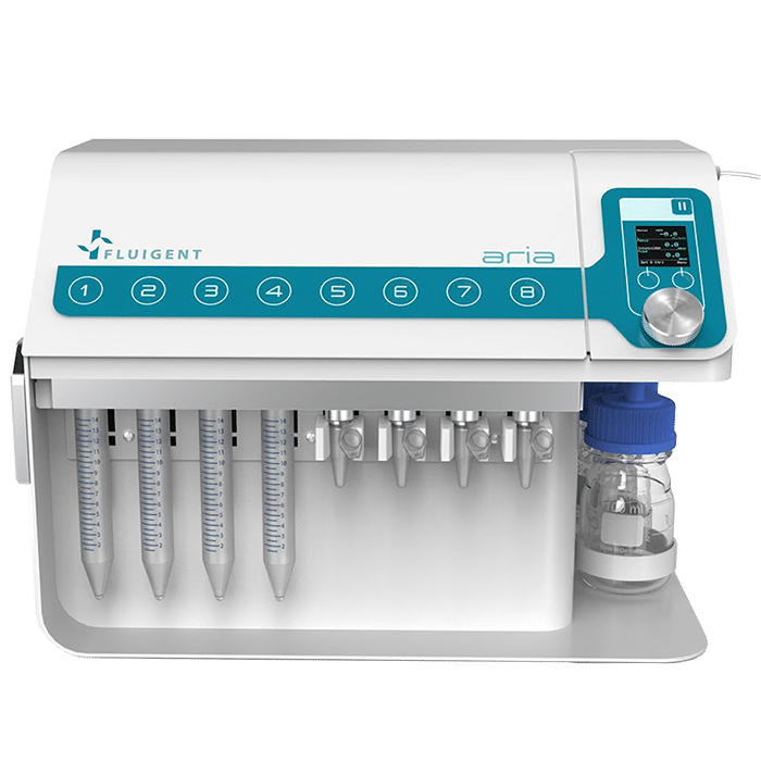
Explore our application note to see how the innovative Fluigent Aria, a software-assisted instrument capable of delivering up to 10 different solutions, automates the entire process of capturing and labeling MDA-MB-231 breast cancer cells.
This includes surface treatment, injection of antibodies-coated beads, cell suspension, and immunostaining. The Aria streamlines cancer cell analysis, saving time, reducing reagent usage, and minimizing error margins.
This study has been made in collaboration with Emile Lakis, post-doctorate at Institut Curie, Team MMBM (Macromolecules and Microsystems in Biology and Medicine)

We have demonstrated the use of Aria and its related software for the automated delivery of different liquids for the capture and labeling of cancer cells (breast cancer) using a complex microfluidic setup. We also demonstrate how Aria can adapt to specific protocols via its software features, allowing for the optimization of protocols.
Key Highlights:
- Automated Cancer Cell Analysis: Discover how the Aria simplifies the capture and labeling of cancer cells, specifically breast cancer, using a sophisticated microfluidic setup.
- Tailored Protocols: See how the Aria adapts to specific protocols through its software features, allowing for protocol optimization.
- Download the Application Note: Access in-depth insights and data.
Understanding Metastasis and Heterogeneity of Circulating Tumor Cells (CTCs)
Cancer, a leading global cause of mortality, is often fatal due to metastasis. Although it begins as a localized disease, it can become systemic before clinical symptoms arise and traditional imaging methods can detect it. Emerging evidence suggests that cancer cells enter the bloodstream long before symptoms manifest, potentially forming distant metastases.
Indeed, it has often become systemic by the time a patient becomes symptomatic and the disease is detected by currently available imaging modalities such as traditional radiography (X-ray), magnetic resonance imaging (MRI), computed tomography (CT), positron emission tomography (PET), or ultrasound. There is growing evidence that cancer cells are shed from the primary tumor into the circulation prior to the presentation of clinical symptoms. These circulating tumor cells (CTCs) may finally colonize at distant sites and form metastases.
Since CTCs are mainly characterized by their morphology and immunostaining pattern, their heterogeneity is a major obstacle for CTC detection. The circulating tumor cells derived from different types of tissues significantly distinguish from each other with different size, shape, and immunophenotyping profiles. However, there is broad morphological and immunophenotypical variation within CTCs derived from the same tissue of origin.
Automating Cancer Cell Analysis for Enhanced CTC Detection and Characterization
Traditional CTC preparation and antibody staining methods are time-consuming and labor-intensive, often requiring exposure to multiple fluids and consuming several hours. This process can involve over five consecutive solutions, demanding precision and controlled flow rates.
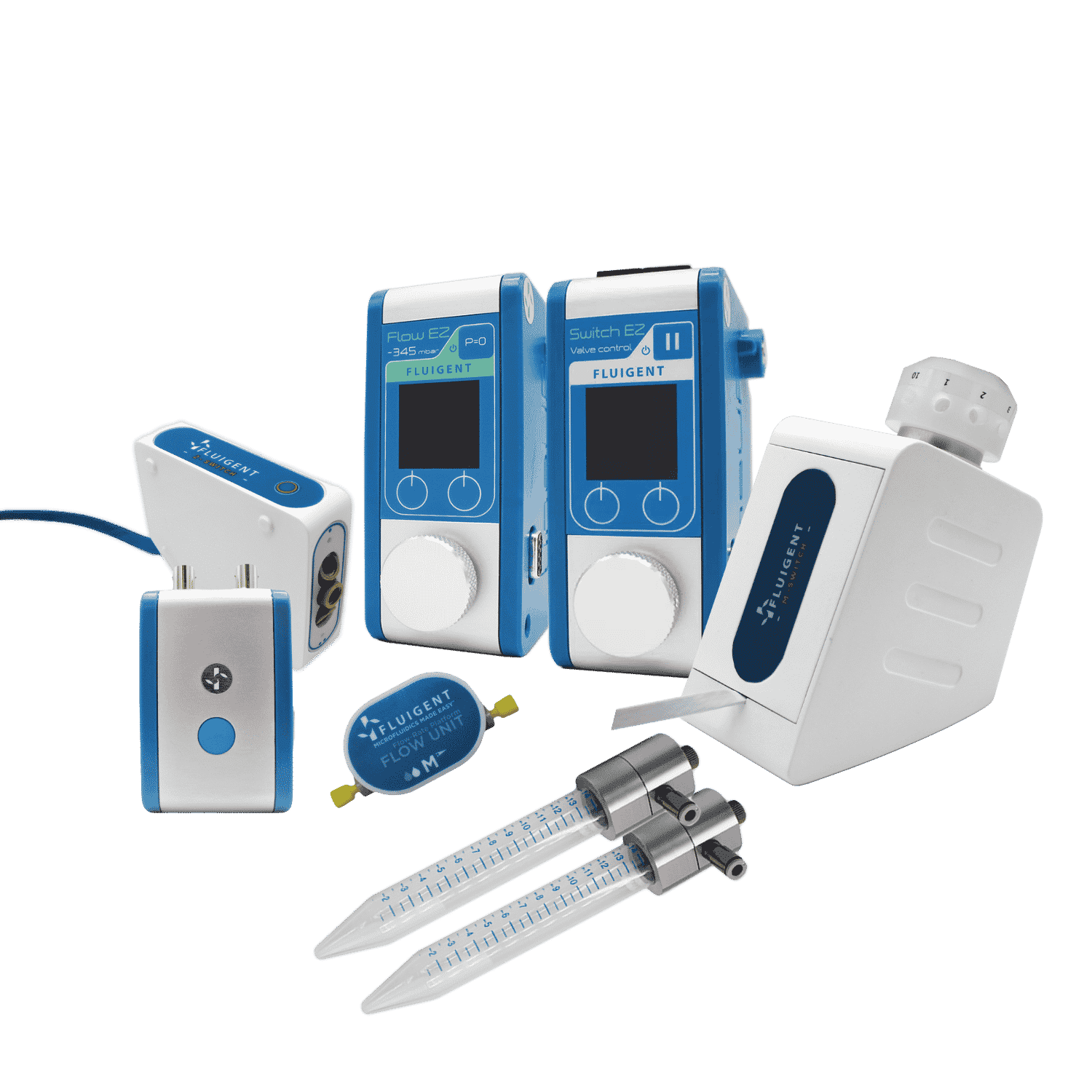
Aria was designed to address these challenges and automate cancer cell analysis by automating fluid supply procedures, ensuring accurate injection volumes and controlled flow rates (ranging from 1 to 1000 µL/min). By maintaining smooth fluid control, it prevents shear stress from altering cell characteristics and damaging samples.
Benefits of Aria: Precise volume control for enhanced analysis
- Streamlined Workflow: Achieve reproducible investigations with automated cell labeling and manipulation, ideal for drug screening and perfusion studies.
- Precise Volume Control: Aria’s capabilities ensure the exact amount of fluid is delivered for enhanced analysis.
Unlocking the Potential of Aria:
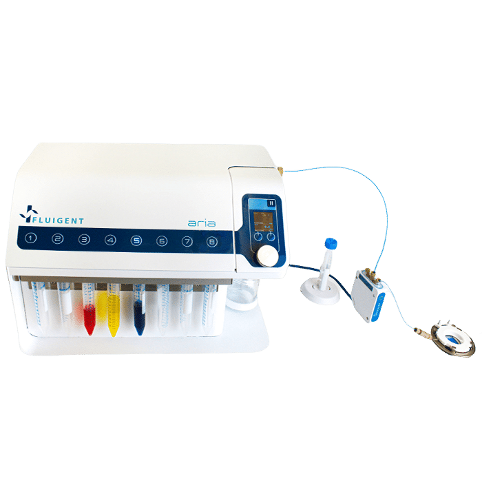
Our application note walks you through a comprehensive, multi-step protocol, illustrating how Aria and its automation software simplify cell capture and detection. We use the EPHESIA microfluidic chip from Institut Curie and the MDA-MB-231 breast cancer cell line to demonstrate the functionality and advantages of our system.
Webinar – Automated fluid delivery for cell and tissue imaging
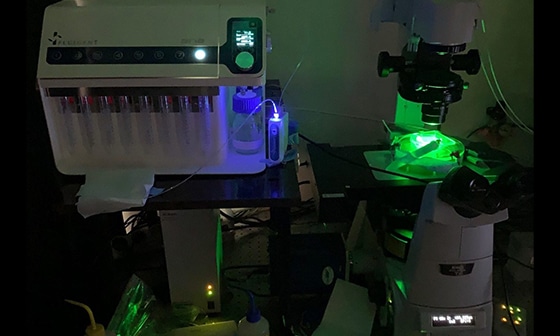
You wish to gain time, precision and reproducibility with your immunofluorescence assay or any other assay requiring injection of multiple solutions on your sample. Follow our webinar to discover how to interface a flow chamber (or a microfluidic chip) to our automated fluid delivery device ARIA. This will allow you to deliver up to 10 different solutions in a sequential and autonomous manner. In addition, ARIA can be synchronized with any microscope to launch an image acquisition cycle and to resume the perfusion protocol once the imaging cycle has been completed.
This all-in-one workflow facilitates the succession of cycles of injection/incubation time with reagents and image acquisition, a feature particularly well adapted to complex cell and tissue imaging. Here, we will present some examples of applications such as multiplexed tissue imaging, DNA-PAINT or seqFISH as well as cell capture and staining.
Planning speech:
Reasons & Advantages to use automated sequential fluid deliver
Demo video
Example of applications:
- Automated multiplexed tissue imaging
- Sequential fluorescence in situ hybridization (SeqFISH)
- Capture and characterization of circulating tumor cells
Q&A session
Conclusion
Aria and its related software were used for the automated delivery of different liquids for the capture and labelling of breast cancer cells using a complex microfluidic setup. This protocol included 10 different liquid injections, surface treatment, beads injection, cells injection, capturing, and labelling in a sequential and automated manner. Thus, we have demonstrated the use of Aria for optimal automated cancer cell analysis.
Aria can also adapt to specific protocols by making use of its specific software features, allowing for the optimization of complex protocols.
Aria provides key features for performing this application as it brings to the user:
- Reduced manipulation time and timed exposure to antibodies, fluorophores, and DNA probes with automated and timed protocols
- Reduced handling for minimal contamination< or changes to cell conditions
- Higher reproducibility compared to manual methods, resulting in more reliable results
References
- Autebert, J. et al. 2015 High purity microfluidic sorting and analysis of circulating tumor cells: towards routine mutation detection. Lab Chip 15, 2090–2101 (2015).
- Kim, M. Y. et al. Tumor Self-Seeding by Circulating Cancer Cells. Cell 139, 1315–1326 (2009).
- Cabel, L. et al. Clinical potential of circulating tumour DNA in patients receiving anticancer immunotherapy. Nat. Rev. Clin. Oncol. 15, 639–650 (2018).
- Bidard, F. C. et al. Clinical validity of circulating tumour cells in patients with me- tastatic breast cancer: A pooled analysis of individual patient data. Lancet Oncol. 15, 406–414 (2014).
- Saias, L., Autebert, J., Malaquin, L. & Viovy, J.-L. 2011 Design, modeling and characte- rization of microfluidic architectures for high flow rate, small footprint microfluidic systems. Lab Chip 11, 822–32 (2011).
- Bernacka-wojcik, I. 2014 Design and development of a microfluidic platform for use with colorimetric gold nanoprobe assays. (Universidade Nova de Lisboa, 2014)

WEBINAR: Enhancing Microfluidic Cell immunolabeling with Aria Technology
Discover
WEBINAR- How to turn your fluorescence microscope into a spatial omics platform
Discover

Expertises & Resources
-
Microfluidic Application Notes Automating Neuronal Cell Immunofluorescence in Microfluidic Chips Read more
-
Microfluidics Case Studies University of Rochester: A tissue chip platform for real-time sensing of secreted inflammatory markers using ARIA Read more
-
Microfluidic Application Notes Automated Immunofluorescence using Aria Read more
-
Microfluidics Case Studies Automated immunolabeling to perform highly multiplexed tissue imaging with ARIA Read more
-
Expert Reviews: Basics of Microfluidics High Throughput Single Cell Analysis Read more