Perfusion Bioreactor for 3D Bioprinted Tissue Models: Dynamic Culture of Skeletal Muscle
Researchers from the University of Bayreuth, FAU Erlangen-Nürnberg, the University of Hohenheim and Fraunhofer IPA built an additively manufactured perfusion bioreactor that combines laminar perfusion, electrical stimulation, in-line pH and temperature sensing and fluorescence microscopy. In 3-day cultures of C2C12 myoblasts, perfusion supported a significant recovery of cell viability in one of three bioinks compared with static controls, and the closed perfusion loop ran on Fluigent instruments (L. Sprenger et al., 2026) [1].
This case study covers perfusion bioreactors for tissue engineering and biofabrication research, operated at microlitre-per-minute flow rates, rather than the large-scale perfusion bioreactors used in biopharmaceutical production.
Key takeaways
- A perfusion bioreactor continuously flows fresh medium through a tissue construct; this 3D-printed design adds electrical stimulation, in-line pH and temperature sensing and microscopy in one system [1].
- A Fluigent MFCS-EX pressure controller drove the closed loop at 80 µL/min, with two Flow Unit M sensors measuring the flow on each side of the chamber.
- The flow direction alternated every 6 hours over 3 days of culture, and in-line sensors recorded stable conditions at 37 °C and pH 7.2.
- In ADA-GEL-MBGN, C2C12 viability recovered to 85.7% after 3 days of perfusion versus 70.5% in static culture; GelMA kept viability above 95% under both conditions.
- Platinum-coated copper electrodes 12 mm apart deliver 0.83–83.33 V/cm for 1–100 V, and isolated murine muscle fibers twitched once the field exceeded 16.67 V/cm.
A Paper from the University of Bayreuth
Paper: Sprenger, L.; Schorzmann, J.; Lu, H.-H.; Osama, M.; Bauer, J.; Haug, M.; Friedrich, O.; Boccaccini, A. R.; Döpper, F.; Salehi, S. Additive-Manufactured, Multifunctional Bioreactor Technology for Dynamic Culture of 3D Bioprinted Tissue Models. Adv. Mater. Technol. 2026, 11, e02635.
This study is the result of a collaboration between Friedrich-Alexander-Universität Erlangen-Nürnberg, the Fraunhofer Institute for Manufacturing Engineering and Automation IPA, the Chair for Manufacturing and Remanufacturing Technology, and the Biomaterials for Tissue Regeneration group at the University of Bayreuth [1,2]. Led by Prof. Dr. Sahar Salehi-Müller, the group combines biomaterials and microtechnologies to engineer functional skeletal muscle tissue [3]. Since October 2025, she has held a joint professorship in the engineering of biointelligent systems at the University of Hohenheim and Fraunhofer IPA in Stuttgart, where the lab is now based [2].
What is a perfusion bioreactor and why does dynamic culture matter?
A perfusion bioreactor is a culture device that continuously flows fresh medium through or around a tissue construct. The flow supplies nutrients and removes metabolic waste products, instead of leaving the construct in still medium that is exchanged by hand [4]. Dynamic culture of this kind supports tissue maturation after biofabrication [4].
In practice, a perfusion bioreactor is a closed loop: a scaffold chamber, media reservoirs and a pump or pressure source that drives nutrient and gas exchange [1]. Bioreactors can also apply mechanical or electrical stimuli, which helps them mimic the in vivo environment and support functional tissue formation [1].
The same principle drives microfluidic cell culture, where a controlled flow of medium replaces periodic manual exchange. Microfluidic technologies are increasingly applied to bioreactor design, as reviewed by Chen et al. (Figure 1) [5].
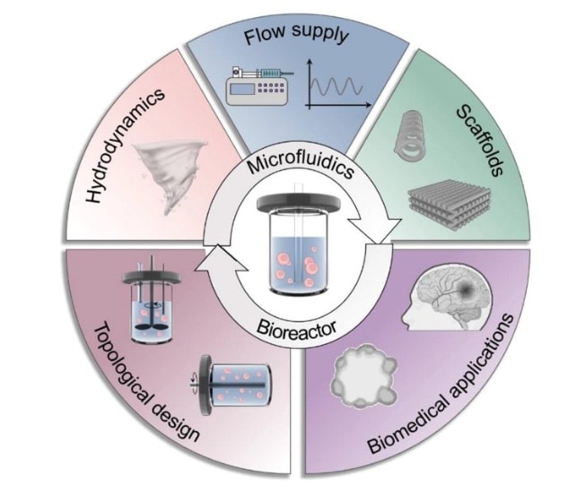
Figure 1. Graphical abstract of the review “Emerging microfluidics for bioreactor”. Reprinted from Chen et al. (2025) [5].
Perfusion bioreactor systems in 3D bioprinting tissue engineering
Additive manufacturing now produces the perfusion bioreactor itself, not only the tissue inside it. According to the authors, it brings geometric freedom, functional integration and multi-material capability that machined commercial systems lack, so the chamber, fluid connections and sensors can be shaped to the tissue model [1,4].
In earlier work involving the same group, Gensler et al. bioprinted perfusable tissue directly into a 3D-printed bioreactor whose geometry could be adapted to a wound-specific shape [4]. The Bayreuth bioreactor follows the same logic: its culture chamber can be resized to the scaffold in rapid design–build–test cycles [1].
The engineering challenge: feeding a 3D bioprinted tissue
Thick constructs cannot rely on diffusion alone. Oxygen and nutrient supply within a scaffold is limited to a diffusion distance of about 150–200 µm; beyond it, engineered tissue requires vascularization to avoid hypoxia-induced cell damage or necrosis [1,6]. One established answer is to perfuse medium directly through the scaffold, which removes the need for manual media changes and supports cell distribution, spreading and viability [1].
Flow alone is not the whole problem. Most current bioreactors focus on medium perfusion and on mechanical or electrical stimulation, but often lack real-time monitoring, which is essential for quality control [1]. According to the authors, no bioreactor prototype combining sensing, electrical stimulation, perfusion and in-line microscopic monitoring had been experimentally validated and published before this work [1].
The Bayreuth team set out to close that gap with one additively manufactured system for skeletal muscle tissue [1].
Aim of the study
The study aimed to build one additively manufactured bioreactor that perfuses, stimulates and monitors a skeletal muscle tissue model, then to test it with living cells against static culture [1].
- Build a multifunctional bioreactor by additive manufacturing, integrating laminar perfusion, electrical stimulation, in-line pH and temperature sensing and microscopy in one system, following the VDI 2221 design guideline [1].
- Culture C2C12 myoblasts in three bioinks: two extrusion-bioprinted (ADA-GEL60 and ADA-GEL-MBGN) and one cast (GelMA) [1].
- Compare dynamic and static culture, measuring cell viability, cell spreading and myotube formation in the bioreactor against Petri-dish controls [1].
- Validate electrical stimulation by calculating, simulating and measuring the electric field, then stimulating isolated murine muscle fibers as a proof of principle [1].
Figure 2 summarises the study design.

Figure 2. Project overview of the study design. Reproduced from Sprenger et al. (2026), Figure 1 [1], CC BY.
Using the MFCS-EX pressure controller for dynamic culture of bioprinted muscle tissue
The setup has two halves. A 3D-printed bioreactor holds the construct, the electrodes and the optical windows; a Fluigent closed-loop circuit drives and measures the medium flowing through it. The two halves meet at M6-threaded fittings set into the chamber walls [1].
Building the bioreactor and the 3D bioprinted constructs
The final design, prototype 5 of five, was printed as a single piece in translucent high-temperature polylactic acid (HT-PLA) on a Bambu Lab X1 Carbon printer [1]. In earlier screening, the team had shown HT-PLA to be non-cytotoxic under DIN EN ISO 10993-5 and resistant to ethanol under DIN EN ISO 175, although autoclaving deforms it [1,7].
Glass slides (42 × 2 mm) sealed with FDA-compliant silicone rings close the chamber at the top and bottom, so a microscope objective can image the whole scaffold from below at a 4.8 mm working distance, including z-stacks [1].
Screw-on caps make the scaffold platform interchangeable, so constructs can be removed for staining or RNA and protein extraction [1]. The total height is limited to 55 mm, so the bioprinter nozzle can reach the glass and print directly in the chamber [1].
The T-shaped electrodes (26 mm long, 6 mm high) were cut from copper by wire electrical discharge machining, then coated with nickel, a palladium–nickel interlayer and a 0.5 µm platinum top layer [1]. The team compared capacitive coupling, with an HT-PLA wall shielding the electrodes from the medium, against direct conductive coupling [1].
In the final design, the culture chamber was set to 12 mm and the HT-PLA wall between electrodes and chamber was removed, so the electrodes stimulate the medium by direct conduction (Figure 3) [1].

Figure 3. Conductive electrode arrangement of the final bioreactor. Adapted from Sprenger et al. (2026), Figure 2 [1], CC BY.
Three bioinks carried C2C12 myoblasts [1]:
- ADA-GEL60, alginate dialdehyde (2.5% w/v) with gelatin (3.75% w/v), cross-linked with 60 mM CaCl₂ and microbial transglutaminase; 1 × 10⁶ cells/mL. The group had earlier found 60 mM CaCl₂ optimal for this formulation, giving a compression modulus of 11.5 kPa, close to native skeletal muscle [8];
- ADA-GEL-MBGN, the same base loaded with 0.1% w/v Zn-doped mesoporous bioactive glass nanoparticles and cross-linked with 100 mM CaCl₂; 1 × 10⁶ cells/mL;
- GelMA, 3.75% w/v gelatin methacrylate photo-cross-linked at 405 nm; 0.5 × 10⁶ cells/mL.
The two ADA-GEL bioinks were extrusion-printed as three 1.5 cm strands on a round glass slide placed in the chamber, and GelMA was cast into the chamber; matching samples in a Petri dish served as static controls [1].
Assembling the fluidic circuit with Fluigent products
The perfusion loop was built from Fluigent pressure-based flow control. An MFCS-EX pressure controller drives the medium from the media reservoirs into the chamber, and one MFCS-EX allowed two bioreactors to run simultaneously [1]. The MFCS-EX offers up to eight independent pressure channels, and because the liquid only contacts air, the instrument never touches the medium [9].
Two M-size Flow Unit flow sensors, one on each side of the chamber, measure the flow in real time and link the media reservoirs to the bioreactor. Together with the MFCS-EX, they form a closed-loop perfusion system run from Fluigent’s A-i-o 2024 software, version 2.3.2 (the previous version of Fluigent’s OxyGEN control software) [1].
An EasyFerm Bio Arc pH sensor (Hamilton) with integrated temperature sensing measures the culture in line. It sits in an airtight 3D-printed reservoir that is continuously perfused with medium leaving the bioreactor, and records 20 values per minute [1].
The software displays both parameters in real time and can raise an alarm if pH deviates by 0.2 from its initial value or temperature exceeds 37.5 °C; it does not apply automated feedback [1]. Figure 4 shows the setup.
The operating parameters were set as follows [1]:
| Parameter | Value |
|---|---|
| Pressure source | MFCS-EX pressure controller |
| Flow sensors | 2 × Flow Unit M, one on each side of the chamber |
| Flow rate | 80 µL/min |
| Flow direction | Alternated every 6 h |
| Bioreactors run in parallel | 2 |
| Pre-culture validation | 72 h of continuous operation in an incubator with pre-warmed DMEM, to detect external leakage |
| Culture duration | 3 days of perfusion |
The slow 80 µL/min flow was chosen to minimise turbulence and scaffold detachment, and alternating its direction every 6 hours ensures homogeneous nutrient perfusion and removal of metabolic byproducts throughout the constructs [1].
This set point sits at the upper edge of the Flow Unit M range, 0 ± 80 µL/min for water; the sensor is bidirectional and temperature-compensated between 10 °C and 50 °C, which suits operation in an incubator [10].

Figure 4. Perfusion and monitoring setup, with temperature and pH recorded on days 1 and 3. Adapted from Sprenger et al. (2026), Figure 4A–C [1], CC BY.
Key results
The results cover three questions: how perfusion changes cell viability and spreading, how it changes myotube formation, and whether the electrodes deliver a usable electric field. Sensor recordings confirmed stable culture conditions at 37 °C and pH 7.2 over the 3 days of culture [1].
Perfusion versus static culture: viability, spreading and differentiation
The clearest effect of perfusion appeared in ADA-GEL-MBGN. Its initial viability of 70.20 ± 6.60% recovered significantly to 85.70 ± 1.90% after 3 days in the bioreactor, whereas it stayed at 70.50 ± 3.14% in static Petri-dish culture (Figure 5) [1]. The authors suggest the lower starting viability may stem from the higher CaCl₂ concentration used for cross-linking [1].
In the other two bioinks, viability was similar under both conditions. GelMA stayed above 95%, while ADA-GEL60 fell from 97.60 ± 0.85% to 83.30 ± 1.57% in the bioreactor and to 81.40 ± 1.70% in static culture [1].
Spreading was scored by cell roundness, from 1 for a perfectly round cell to 0 for a fully spread one [1]. Cells spread markedly in all three bioinks over 3 days, but roundness at day 3 did not differ significantly between bioreactor and Petri dish [1]. The authors note that 3 days of culture was not yet long enough to reveal the advantages of perfusion [1].
| Measure | Day 0 | Static culture, day 3 | Perfusion, day 3 |
|---|---|---|---|
| GelMA viability | 95.00 ± 1.34% | 98.00 ± 1.70% | 96.00 ± 1.70% |
| ADA-GEL60 viability | 97.60 ± 0.85% | 81.40 ± 1.70% | 83.30 ± 1.57% |
| ADA-GEL-MBGN viability | 70.20 ± 6.60% | 70.50 ± 3.14% | 85.70 ± 1.90% |
| GelMA roundness | 0.86 ± 0.03 | 0.59 ± 0.03 | 0.53 ± 0.07 |
| ADA-GEL60 roundness | 0.80 ± 0.02 | 0.60 ± 0.06 | 0.60 ± 0.06 |
| ADA-GEL-MBGN roundness | 0.80 ± 0.04 | 0.60 ± 0.05 | 0.50 ± 0.08 |

Figure 5. Cell roundness and viability at day 0 and after 3 days in the bioreactor or a Petri dish. Adapted from Sprenger et al. (2026), Figure 5A, B,C [1], CC BY.
Differentiation was tested on GelMA films: cells grew for 2 days, then differentiated for 3 days with a 24-hour ProFuse S1 supplementation, under perfusion or in a Petri dish [1].
Under perfusion, myotubes reached 283 ± 166 µm in length and 61 ± 13.4 µm in width, against 240 ± 140.40 µm and 58 ± 18.30 µm in static culture; only the width difference reached significance (p = 0.05), and the fusion index was similar, 12.00 ± 3.98% versus 11.50 ± 1.40% [1].
Alignment matters for muscle models in particular. Skeletal muscle is an anisotropic tissue, and the group’s earlier bioink work aimed specifically at constructs whose cells line up in one direction [8]. The paper’s abstract reports faster myoblast alignment in ADA-GEL after 3 days of perfusion, and the authors name the effect of flow conditions on alignment as a priority for future studies [1].
Electrical stimulation validated in the same chamber
The capacitive configuration tested in earlier prototypes produced only micro- to sub-millivolt fields, several orders of magnitude below the more than 4–6 V/cm reported as physiologically relevant for myotubes. The team therefore switched to direct conductive stimulation in the final bioreactor [1].
In the conductive configuration, the field follows from the applied voltage and the electrode spacing: E = V / d, where E is the electric field (V/cm), V the applied voltage amplitude (V) and d the electrode distance (cm) [1].
As a proof of principle, single intact fibers of murine Mm. interossei plantares were placed in the chamber and stimulated with 2 ms square-wave pulses at 1 Hz for 20 s, stepping from 8.33 to 83.33 V/cm [1].
Conclusion
Sprenger et al. show that a perfusion bioreactor built by additive manufacturing can perfuse, stimulate and monitor a skeletal muscle tissue model in a single system. Perfusion supported a significant viability recovery in ADA-GEL-MBGN, 85.7% versus 70.5% in static culture after 3 days, and the conductive electrodes made isolated muscle fibers twitch from 16.67 V/cm [1].
The platform’s value lies in combining functions that usually need separate setups. A printed, resizable housing carries the electrodes and optical windows, while standard Fluigent flow control runs the closed perfusion loop and reports the flow on each side of the chamber [1]. The authors plan longer cultures and varied flow conditions to study myoblast alignment and the combined effects of perfusion and electrical stimulation [1].
Read the full open-access article: Additive-Manufactured, Multifunctional Bioreactor Technology for Dynamic Culture of 3D Bioprinted Tissue Models [1].
Planning a perfusion setup for your own tissue constructs? Contact us to discuss your flow rates, chamber and monitoring needs with our application team.
Related products



Related Expertises
-
Expert Reviews: Basics of Microfluidics What Is Organ-on-a-Chip? A Complete Guide Read more
-
Microfluidics Case Studies CNRS/UTC: study of a liver-on-a-chip model Read more
-
Expert Reviews: Basics of Microfluidics Fluid Shear Stress in Cell Biology: Why and How to Control It Read more
-
Microfluidic Application Notes Cartilage-on-a-chip, an example of complex mechanical stimulation using Fluigent’s technology Read more
-
Expert Reviews: Basics of Microfluidics Passive and active mechanical stimulation in microfluidic systems Read more
References
- Sprenger, L., Schorzmann, J., Lu, H.-H., Osama, M., Bauer, J., Haug, M., Friedrich, O., Boccaccini, A. R., Döpper, F., & Salehi, S. (2026). Additive-manufactured, multifunctional bioreactor technology for dynamic culture of 3D bioprinted tissue models. Advanced Materials Technologies, 11(17), e02635. https://doi.org/10.1002/admt.202502635
- University of Hohenheim. (2026). Prof. Dr. Sahar Salehi-Müller. https://salehilab.uni-hohenheim.de/en/profdrsaharsalehi-mller
- University of Hohenheim, Department of Biointelligent Systems Engineering. (2026). Development of printable and cell compatible bioink for 3D bioprinting of skeletal muscle tissue. https://salehilab.uni-hohenheim.de/en/3dbioprint
- Gensler, M., Malkmus, C., Ockermann, P., Möllmann, M., Hahn, L., Salehi, S., Luxenhofer, R., Boccaccini, A. R., & Hansmann, J. (2024). Perfusable tissue bioprinted into a 3D-printed tailored bioreactor system. Bioengineering, 11(1), 68. https://www.mdpi.com/2306-5354/11/1/68
- Chen, H., Fang, Y., Luo, Z., Wang, Y., Sun, W., & Zhao, Y. (2025). Emerging microfluidics for bioreactor. Materials Today, 86, 356–392. https://doi.org/10.1016/j.mattod.2025.03.009
- Folkman, J., & Hochberg, M. (1973). Self-regulation of growth in three dimensions. The Journal of Experimental Medicine, 138(4), 745–753. https://doi.org/10.1084/jem.138.4.745
- Schorzmann, J., Gerstl, H., Tan, Z., Sprenger, L., Lu, H.-H., Taumann, S., Wimmer, M., Boccaccini, A., Salehi, S., & Döpper, F. (2023). Materials screening methodology for additive manufacturing in bioreactor technology. In Solid Freeform Fabrication 2023: Proceedings of the 34th Annual International Solid Freeform Fabrication Symposium (pp. 2006–2028). University of Texas at Austin. https://doi.org/10.26153/tsw/51099
- Sprenger, L., Lu, H.-H., Trippmacher, S., Mansfeld, U., Milkin, P., Ionov, L., Papastavrou, G., Boccaccini, A. R., & Salehi, S. (2024). Composite alginate dialdehyde-gelatin (ADA-GEL) hydrogel containing short ribbon-shaped fillers for skeletal muscle tissue biofabrication. ACS Applied Materials & Interfaces, 16(34), 44605–44622. https://doi.org/10.1021/acsami.4c10751
- Fluigent. (2026). Microfluidic flow control system: MFCS series. https://www.fluigent.com/research/instruments/pressure-flow-controllers/mfcs-series/
- Fluigent. (2026). FLOW UNIT: Bidirectional microfluidic flow sensor. https://www.fluigent.com/research/instruments/sensors/flow-unit/