A Microfluidic Approach for High-Throughput Raman Spectroscopy of Whole Blood
Raman spectroscopy is a powerful tool for label-free biochemical analysis, but its application to complex biological fluids such as whole blood remains limited by low signal intensity, long acquisition times, and photodamage risks.
To overcome these challenges, F. Zorzi et al. introduced a microfluidic lab-on-a-chip platform enabling 3D hydrodynamic focusing. This approach allows full-volume, low-noise, and multiplexed Raman analysis of non-transparent fluid with rapid acquisition and real-time diagnostics.[1]
Enabled by Fluigent’s pressure-based flow control, it ensures precise and stable hydrodynamic focusing, supporting reproducible conditions, and improved efficiency in microfluidic Raman analysis.
A paper from Istituto Italiano di Tecnologia and Politecnico di Milano
Paper: Zorzi F, Jensen EA, Serhatlioglu M, Bonfadini S, Dziegiel MH, Criante L, et al. Flow cell for high throughput Raman spectroscopy of non-transparent solutions. Lab Chip. 2024 Dec 17;25(1):69–78.
This study was carried out through a collaboration between leading academic and clinical institutions specializing in nanotechnology, microfluidics, and biomedical analysis. The work was led by researchers from the Center for Nano Science and Technology at the Istituto Italiano di Tecnologia (IIT, Milan, Italy), in partnership with the Politecnico di Milano and the Technical University of Denmark (DTU).
The project also involved clinical expertise from Copenhagen University Hospital, enabling access to real human blood samples and ensuring strong relevance for biomedical applications. This interdisciplinary collaboration brought together expertise in microfabrication, optical spectroscopy, and clinical diagnostics, providing anideal framework for developing and validating innovative Raman-based analytical platforms.
Raman Spectroscopy
Raman spectroscopy is an optical technique used to probe the molecular composition of a sample. It relies on the inelastic scattering of light: when a laser interacts with molecules, a small fraction of the scattered light undergoes a shift in energy that is characteristic of the vibrational modes of the molecules. This spectral fingerprint enables the identification and analysis of chemical species without the need for dyes or markers. [2,3]
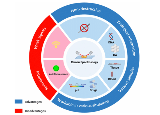
Because of its specificity and non-destructive nature, Raman spectroscopy is widely used across multiple fields, including chemistry, materials science, and biomedical diagnostics. In particular, it has gained strong interest for biological applications such as cell analysis, pathogen detection, and blood diagnostics, where label-free and real-time measurements are highly valuable (Figure 1). [2,3]
However, despite its potential, Raman spectroscopy faces several key limitations. The Raman signal is inherently weak, leading to long acquisition times and limiting throughput. In complex and non-transparent samples, signal attenuation and background noise further degrade performance. [2,3]
Why use Raman spectroscopy in Biology: The Case of Whole Blood
In the context of whole blood, Raman spectroscopy enables the molecular characterization of individual cellular components, including red blood cells, white blood cells, and platelets. Each cell type exhibits a distinct spectral signature, allowing their identification and detailed analysis of biochemical composition. This capability is particularly valuable for investigating cellular heterogeneity and detecting disease-related alterations at the single-cell level.
However, whole blood is a highly complex and challenging matrix for Raman analysis. The strong optical absorption of hemoglobin, combined with high cell density and the presence of plasma proteins, generates significant background interference and signal distortion. In addition, the coexistence of multiple cell populations makes it difficult to selectively probe and analyze individual cells in a reliable and high-throughput manner. These limitations highlight the need for approaches that can precisely control cell positioning and enable continuous, flow-based measurements, paving the way for more robust and scalable analysis. [5,6]
Aim of the study
This study aims to overcome the major limitations currently hindering the broader application of Raman spectroscopy to complex biological samples, such as whole blood. By developing a microfluidic lab-on-a-chip platform, F. Zorzi et al. enhance Raman analysis by increasing throughput, minimizing sample preparation, and enabling evaluation of the entire sample volume. The goal is to provide faster, more efficient, and scalable measurements by leveraging hydrodynamic focusing and flow-based analysis.
Methodology: How to Couple Microfluidics with Raman Spectroscopy
Whole blood samples used in this study were collected from voluntary donors at Copenhagen University Hospital and stored in EDTA tubes at 4°C. During experiments, blood was injected as the sample phase, while a PBS-based buffer containing diluted EDTA was used as a sheath fluid for hydrodynamic focusing.
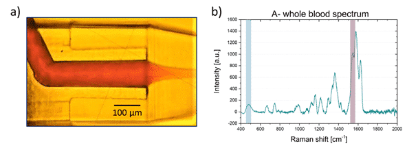
The microfluidic chip was fabricated in fused silica. It employs 3D hydrodynamic focusing to confine and center the sample stream within a surrounding buffer flow, enabling precise control of the stream size (down to 15 µm) and optimal alignment with the Raman excitation volume. A locally thinned observation region enhanced signal collection while reducing background noise. This design also supported parallel flow multiplexing, allowing high-throughput and simultaneous analysis of multiple samples (Figure 2).
To ensure stable and precise flow control,the microfluidic set-up relied on a pressure-driven system composed of a Fluigent MFCS pressure controller which regulates the sample and buffer reservoirs, along with two Fluigent Flow Unit sensors for real-time monitoring of flow rates upstream of the chip. By adjusting the pressure ratio between the sample and sheath flows, the sample stream was hydrodynamically focused at the center of the microchannel.
Raman measurements were performed using a 532 nm continuous-wave laser focused into the microchannel through a high-NA objective, which also collected the backscattered signal. The Raman signal was separated from the excitation light using dichroic and notch filters, then analyzed via an imaging spectrometer coupled to a cooled CCD camera.


Proof of Concept: Microfluidic Raman Analysis of Whole Blood
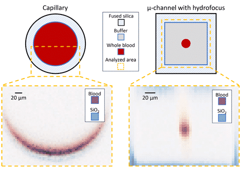
The study showed improved performance enabled by the microfluidic approach compared to conventional capillary-based measurements. As displayed in Figure 3, precise hydrodynamic focusing allowed the sample stream to be confined down to 15 µm and matched to the excitation volume, enabling full-volume interrogation within a single measurement. This resulted in a linear relationship between Raman signal intensity and analyte concentration (Figure 4), confirming the system’s quantitative capability down to micromolar ranges.


When applied to whole blood, the impact of microfluidics becomes more observed. In Figure 5 and Figure 6, the microfluidic chip enabled signal collection from nearly 100% of the flowing sample, whereas capillary-based measurements probe only a small fraction (8%), with significant background contribution from the walls.
This improved sampling efficiency, combined with uniform flow conditions at the channel center, leads to more representative and reproducible measurements, especially for heterogeneous or corpuscular fluids. Therefore, the platform enabled acquisition of Raman spectra to be more representative of the full cell population, which is particularly critical for detecting rare pathological cells.
Importantly, this microfluidic configuration enhances throughput while reducing photodamage. The higher flow velocity increased the number of cells analyzed per unit time, while the reduced interaction time allowed the use of higher excitation powers without inducing degradation. This resulted in stronger signals and shorter acquisition times, demonstrating the clear benefit of microfluidics for high-throughput Raman analysis.
Conclusion
In this study, researchers from the Istituto Italiano di Tecnologia and Politecnico di Milano demonstrated that coupling Raman spectroscopy with a microfluidic lab-on-a-chip platform, enables efficient, label-free analysis of complex fluids such as whole blood. This approach allows complete sample characterization, enhances signal quality, and markedly increases throughput, while minimizing acquisition time and photodamage. Overall, it underscores the potential of integrating advanced microfluidics to Raman spectroscopy to deliver reliable, high-throughput, and clinically relevant diagnostic solutions.
Related Extertises
-
Microfluidics Case Studies Micropipette Aspiration of Red Blood Cells Mechanosensitivity and Mechanics Read more
-
Expert Reviews: Basics of Microfluidics Why Control Shear Stress in Cell Biology? Read more
-
Expert Reviews: Basics of Microfluidics Extended Capabilities of Pressure Driven Flow for Microfluidic Applications Read more
References
- Zorzi F, Jensen EA, Serhatlioglu M, Bonfadini S, Dziegiel MH, Criante L, et al. Flow cell for high throughput Raman spectroscopy of non-transparent solutions. Lab Chip. 2024 Dec 17;25(1):69–78. doi:10.1039/D4LC00586D
- Keresztury G. Raman Spectroscopy: Theory. In: Chalmers JM, Griffiths PR, editors. Handbook of Vibrational Spectroscopy. 1st edn. Wiley; 2001 [cited 2026 Apr 7]. Available from: https://onlinelibrary.wiley.com/doi/10.1002/0470027320.s0109 doi:10.1002/0470027320.s0109
- Kudelski A. Analytical applications of Raman spectroscopy. Talanta. 2008 Jun 30;76(1):1–8. doi:10.1016/j.talanta.2008.02.042
- Li C, Feng C, Xu R, Jiang B, Li L, He Y, et al. The emerging applications and advancements of Raman spectroscopy in pediatric cancers. Front Oncol. 2023 Feb 6;13. doi:10.3389/fonc.2023.1044177
- Jensen EA, Serhatlioglu M, Uyanik C, Hansen AT, Puthusserypady S, Dziegiel MH, et al. Label-Free Blood Typing by Raman Spectroscopy and Artificial Intelligence. Advanced Materials Technologies. 2024;9(2):2301462. doi:10.1002/admt.202301462
- Laskowska P, Mrowka P, Glodkowska-Mrowka E. Raman Spectroscopy as a Research and Diagnostic Tool in Clinical Hematology and Hematooncology. International Journal of Molecular Sciences. 2024 Mar 16;25(6). doi:10.3390/ijms25063376