Breathing Lung-on-Chip Platform for Dynamic Anti-Fibrotic Testing
Researchers from the University of Bern and AlveoliX developed a breathing lung-on-chip model that mimics the dynamic mechanical environment of human alveoli. By combining a collagen-elastin membrane with cyclic stretch, the platform provides a physiologically relevant model of idiopathic pulmonary fibrosis.
The study demonstrates that breathing-like mechanical stimulation significantly influences fibrotic responses and anti-fibrotic drug efficacy.
Advancing research with organs-on-chips technologies
Organ-on-chip technologies have emerged as powerful tools to mimic human physiological environments in vitro, enabling more predictive models for disease research and drug testing than traditional systems. Among these, lung-on-chip platforms are particularly promising, yet accurately reproducing the complex structure and dynamic mechanical behavior of the alveoli remains a significant challenge, especially for the study of idiopathic pulmonary fibrosis.
Accordingly, Weber et al. (2026) developed a microengineered alveolar array lung-on-chip integrating a biologically derived collagen–elastin membrane with controlled cyclic mechanical actuation, enabling physiologically relevant, reproducible breathing-like motions at the microscale.
This platform combines Fluigent’s pressure-based flow control with an innovative array of alveoli made with a suspended collagen-elastin membrane developed by the University of Bern and AlveoliX alveolar epithelial cells to provide precise and stable mechanical stimulation for fibrosis modeling and the assessment of anti-fibrotic drug responses.
Development of a lung-on-chip model, a paper from the University of Bern and AlveoliX
Paper: Weber TA, Zamprogno P, Schneider S, Hajari MA, Büchler P, Hobi N, et al. Microengineered alveolar array lung-on-chip with hydrogel membrane and simulated breathing mechanics for anti-fibrotic testing. Biofabrication. 2026 Jan;18(1):015026.
This study resulted from a collaboration between the ARTORG Center for Biomedical Engineering Research (University of Bern, Switzerland) and AlveoliX, which develops organs-on-chip technology for predictive preclinical research.
Together, these partners bring complementary strengths in biofabrication, microfluidics, and respiratory disease modeling, providing a robust framework for the development and validation of physiologically relevant lung-on-chip technologies.
Pathological context: the Idiopathic Pulmonary Fibrosis
Idiopathic pulmonary fibrosis is a chronic and progressive interstitial lung disease characterized by excessive scarring of the lung tissue, leading to irreversible loss of respiratory function. The disease causes progressive dyspnea, chronic dry cough, fatigue, and reduced exercise tolerance. Idiopathic pulmonary fibrosis mainly affects adults over 60 years old, especially men and former smokers. This severe lung disease has a poor prognosis, with a median survival of 3 to 5 years after diagnosis (Figure 1). [1,2,3]
Current treatment options remain limited. Supportive care, including oxygen therapy and pulmonary rehabilitation, is commonly used to improve quality of life, while lung transplantation remains the only definitive treatment for eligible patients with advanced disease. Anti-fibrotic drugs can slow disease progression, but they do not reverse existing fibrosis or provide a cure. Nowadays, several innovative tools have emerged, offering great opportunities for more predictive and physiologically relevant in vitro models to better understand fibrosis mechanisms and accelerate the development of new therapies. [3,4]

Why use Organ-on-chip for Idiopathic Pulmonary Fibrosis Study?
Organ-on-chip technologies are emerging microphysiological systems designed to recreate key structural and functional features of human organs within miniaturized microfluidic devices. By combining microengineering, biomaterials, and cell biology, these platforms provide controlled microenvironments where human cells can be cultured under biochemical and mechanical conditions (Figure 2). [6,7]
Disease modeling, drug screening, and toxicity assessment can benefit from organ-on-chip technologies. As an example, lung-on-chip models show strong potential for mimicking complex pulmonary physiology and improving the prediction of human drug responses compared to traditional in vitro and animal models. [6,7]

Organ-on-chip technologies have major advantages including reduced reliance on animal experimentation, along with the use of human cells, improved experimental control, real-time monitoring, lower reagent consumption, and compatibility with high-throughput approaches. However, important challenges remain, including reproducing physiologically relevant mechanical environments while ensuring stable and reproducible long-term operation of complex systems. [7]
Replicating the human lung in vitro in a pathological context
This study addresses key limitations of current lung-on-chip models in accurately replicating the biomechanical environment of the human alveoli. Weber et al. developed a microengineered platform that mimics physiologically relevant breathing motions, enabling the study of lung behavior under dynamic mechanical conditions. The system supports investigations of pulmonary fibrosis and drug responses in a controlled in vitro setting.

Methodology: How to Reproduce Breathing Mechanics in a Lung-on-Chip Platform
The lung-on-chip device developed in this study is based on a microengineered architecture integrating a thin, suspended collagen–elastin hydrogel membrane supported by a hexagonal gold mesh, reproducing the size and geometry of native alveoli.
The platform builds on technologies developed by the University of Bern, including AlveoliX AXiAECs human alveolar epithelial cells. The system is fabricated using injection-molded cyclic olefin copolymer layers assembled with a PDMS membrane acting both as a diaphragm and as microfluidic valves. Pneumatic activation generates controlled pressure differentials, inducing reproducible breathing-like deflections of the hydrogel membrane across an array of alveolar units (Figure 3).
As displayed in Figure 4, human alveolar epithelial cells provided by AlveoliX and lung fibroblasts are co-cultured on opposite sides of the membrane to recreate the alveolar barrier. Cells are cultured under static or dynamic conditions, with cyclic mechanical stimulation applied to mimic physiological breathing. Early fibrotic changes are induced via transforming growth factor-beta 1 (TGF-β1) stimulation, while the anti-fibrotic drug nintedanib is used to assess therapeutic effects. Barrier integrity and cellular responses are then analyzed using TEER measurements, immunofluorescence staining, ELISA assays, and gene expression analysis.

To ensure precise and stable control of both fluidic and mechanical conditions, the system relies on pressure-driven flow provided by Fluigent flow control solutions. In this setup, the Flow EZ is used to generate the cyclic pressure (shear stress) required to drive the breathing-like activation of the membrane, delivering controlled oscillatory signals that reproduce physiological respiratory motions. In parallel, the Push Pull controls the on-chip pneumatic valves, enabling precise regulation of medium exchange within the microfluidic network. By independently controlling these two pressure lines, the platform ensures synchronized and reproducible mechanical stimulation and fluid handling.


Proof of Concept: Dynamic Lung-on-Chip Modeling of Anti-Fibrotic Testing
The study demonstrates the ability of the lung-on-chip platform to reproduce physiologically relevant breathing-like mechanical stimulation at the microscale.
As shown in Figure 5, controlled pneumatic actuation induced a stable and reproducible membrane deflection of approximately 43 µm, generating a spatially heterogeneous strain distribution across each alveolar unit, with a maximum surface increase of about 7.8%.
This gradient closely mimics the mechanical environment experienced by cells in native alveoli and was further validated by the upregulation of stretch-responsive markers under cyclic conditions.
When applied to a fibrosis model, the impact of dynamic mechanical stimulation became particularly evident. As illustrated in Figure 6, treatment with TGF-β1 induced extracellular matrix overexpression with significantly stronger effects observed under cyclic stretch compared to static conditions. This highlights the key role of breathing-like forces in amplifying fibrotic signaling and better reproducing disease progression in vitro.


Importantly, the platform also enabled the evaluation of therapeutic responses under physiologically relevant conditions. As shown in Figure 7, the anti-fibrotic drug demonstrates limited efficacy under static culture but significantly improved barrier integrity with enhanced tight junction formation (ZO-1) and reduced fibrotic marker expression under dynamic conditions. These results underscore the critical role of mechanical cues in modulating drug response and confirm the relevance of the system as a predictive tool for preclinical testing.
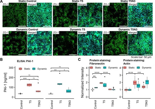
Figure 7: Effect of the anti-fibrotic drug nintedanib on the progression of IPF-like symptoms. (A) Immunofluorescence staining of alveolar epithelial cells for the tight junction protein Zonula Occludens-1 (ZO-1) (green) and nuclei (cyan). (B) Secretion of PAI-1 in static and dynamic culture, treated with TGF-β1 (T5), in combination with nintedanib (T5N3), and control (CTRL) in the co-culture model. (C) Normalized intensity of the fluorescence staining of actin and fibronectin in a fibroblast monoculture.[1]
Lung-on-Chip: A Powerful Tool for Disease Modeling and Drug Discovery
In this study, researchers from the University of Bern and AlveoliX demonstrated that a microengineered alveolar lung-on-chip integrating a collagen-elastin membrane with breathing-like mechanical stimulation enables a more physiologically relevant model of idiopathic pulmonary fibrosis. The platform successfully reproduces dynamic alveolar deformation and reveals the major impact of cyclic stretch on fibrotic responses and drug efficacy.
Enabled by Fluigent’s pressure-based flow control technologies, the system ensures stable and reproducible mechanical stimulation and fluid handling. Overall, this work highlights the potential of advanced lung-on-chip platforms to improve preclinical anti-fibrotic drug testing and support the development of more predictive in vitro pulmonary disease models.
Related Resources
-
Microfluidics Case Studies A Microfluidic Approach for High-Throughput Raman Spectroscopy of Whole Blood Read more
-
Expert Reviews: Basics of Microfluidics Organ-on-chip Platforms in Modern Drug Development and Testing Read more
-
Expert Reviews: Basics of Microfluidics 5 Key Tips for Starting Organ-on-Chip Models Read more
-
Microfluidics White Papers A review of Organ on Chip Technology – A White Paper Read more
-
Expert Reviews: Basics of Microfluidics Why Control Shear Stress in Cell Biology? Read more
-
Expert Reviews: Basics of Microfluidics Mimicking in-vivo environments: biochemical and biomechanical stimulation Read more
References
- Weber TA, Zamprogno P, Schneider S, Hajari MA, Büchler P, Hobi N, et al. Microengineered alveolar array lung-on-chip with hydrogel membrane and simulated breathing mechanics for anti-fibrotic testing. Biofabrication. 2026 Jan;18(1):015026. doi:10.1088/1758-5090/ae2e44
- Meltzer EB, Noble PW. Idiopathic pulmonary fibrosis. Orphanet J Rare Dis. 2008 Mar 26;3(1):8. doi:10.1186/1750-1172-3-8
- Gross TJ, Hunninghake GW. Idiopathic Pulmonary Fibrosis. New England Journal of Medicine. 2001 Aug 16;345(7):517–25. doi:10.1056/NEJMra003200
- Barratt SL, Creamer A, Hayton C, Chaudhuri N. Idiopathic Pulmonary Fibrosis (IPF): An Overview. Journal of Clinical Medicine. 2018 Aug 6;7(8). doi:10.3390/jcm7080201
- Idiopathic Pulmonary Fibrosis | Pulmonary Fibrosis Foundation. Available from: https://www.pulmonaryfibrosis.org/understanding-pff/types-of-pulmonary-fibrosis/idiopathic-pulmonary-fibrosis
- Mittal R, Woo FW, Castro CS, Cohen MA, Karanxha J, Mittal J, et al. Organ-on-chip models: Implications in drug discovery and clinical applications. Journal of Cellular Physiology. 2019;234(6):8352–80. doi:10.1002/jcp.27729
- Srivastava SK, Foo GW, Aggarwal N, Chang MW. Organ-on-chip technology: Opportunities and challenges. Biotechnology Notes. 2024 Jan 1;5:8–12. doi:10.1016/j.biotno.2024.01.001