• 简体中文
  • 한국어
  • Deutsch
Fluigent
  • Research
      • Flow EZ™ microfluidic flow controller
      • Microfluidic Research Equipment
      • Microfluidic Instruments
        • Omi, an Automated Organ-On-A-Chip Platform
        • Automated Perfusion System for Spatial Omics 
        • Microfluidic Pressure Based Flow Controller
        • Microfluidic Sensors
        • Microfluidic Valves
        • Pressure Control Reservoirs
        • Microfluidic Pressure Sources
        • Microfluidic Chips
        • Microfluidic Accessories
      • Software Solutions
        • Lab Integration Software
        • Real-Time Control & Lab Automation Software 
      • Microfluidic Packs
        • UV-crosslinked microcapsule production platform 
        • Encapsulation Platform for FACS
        • Microfluidic Complex Emulsion Production Platform
        • Microfluidic Application Packs
        • Microfluidic Starter Packs
      • Research Applications
        • Microfluidics for Cell Analysis
        • Microfluidics for Organ-on-chip Cell culture
        • Microfluidics for Droplet Generation
        • Microfluidics for Cell Biology
      • Subscribe to the newsletter
  • Industrial
      • Microfluidic OEM
      • Microfluidic OEM Devices
        • Microfluidic OEM Components
        • Customizable OEM Flow Control Modules
      • Fully Custom Microfluidic Device
      • Custom microfluidic device
      • Technologies
        • A Microfluidic Pressure Controller Comparison for Your Ultimate Fluid Control System
        • 5 reasons to choose OEM pressure controllers over OEM syringe pumps for microfluidic applications
        • Microfluidic recirculation system 
        • DFC, “Self-Learning” Microfluidic Flow Control Algorithm
        • Non-Intrusive Flow Sensing Technology
        • Compact All-In-One Microfluidic Micropump
        • Liquid Stirring Solutions
      • Industrial Applications
        • Combining Microfluidics and Spectroscopy
        • Valve Automation with the F-OEM for Microfluidic Applications
        • Localization microscopy and flow control for multiplexing 
        • Contamination-free Liquid Handling System
        • Microfluidic Drug Discovery 
        • Flow Expertise for Cell Encapsulation and Single-Cell Analysis
        • Droplet Digital PCR (ddPCR)
      • Subscribe to the newsletter
  • Markets & Applications
    • Microfluidics in Life Science
    • Microfluidics for Pharmaceutical Applications
    • Microfluidics for Food testing & Agriculture
    • Microfluidics in Cosmetics
    • Microfluidics in Water analysis
  • Company
    • About us
    • Fluigent’s Academic Partners
      • Scientific Partners
      • Fluigent’s Brand Ambassadors
      • Center Partners
    • Team
    • News
    • Events & Webinars
    • Fluigent Newsletter
    • Fluigent’s Distributors
    • Careers
  • Resources & Support
      • Expert Reviews: Basics of Microfluidics
        • General overview of microfluidics
        • Advantages of pressure-based microfluidics
        • Microfluidics tips
        • Droplet & Particle Generation
        • Microfluidic cell biology
        • Industrial / OEM Expertise
        • Funded research program
      • FAQ
      • Videos
        • Expertise videos
        • Product presentation videos
        • Tutorial videos
      • Expertise
        • Videos
        • Microfluidics Article Reviews
        • Microfluidic Application Notes
        • Microfluidics case studies
        • Interviews & Testimonials
        • Microfluidics White Papers
      • Documentation
        • CAD
        • Fluigent Catalog
        • Fluigent products manual
        • Fluigent Products Datasheets
        • Safety datasheet
        • Fluigent Media Kit product icons & images
      • Microfluidic Calculators
        • Shear Stress Calculator
        • Pressure & Flow Rate Calculator
        • Droplet Size Calculator
      • Download software
        • FEZ and Link Firmware Updater​
        • OxyGEN
        • Software Development Kit
        • Discontinued software
      • Subscribe to the newsletter
  • Webshop
  • Contact us
Home » Resources » Expertise » Expert Reviews: Basics of Microfluidics » Funded research program » MYOCHIP | H2020 European project

MYOCHIP | H2020 European project

Introduction

Myochip: Building a 3D innervated and irrigated muscle on a chip

The aim of the European Union’s Horizon H2020 project, Myochip  (https://myochip.imm.medicina.ulisboa.pt) is to build a muscle on chip. Myochip gathers 4 partners from all around the world: The institute of molecular medicine (Portugal), The University of Edinburg (Scotland), and Institut Curie (France).

Seeding cells in hydrogels: the benefit of pressure based flow controllers

Organs on chips are considered as the next generation in vitro models to replace traditional 2D cell culture both in fundamental science and in drug development. This technology focuses on reproducing the microenvironment of cells at the cellular level:

  • The intercellular interaction by positioning one cell type relative to another
  • The extracellular matrix by reproducing both its composition and its microarchitecture
  • The local mechanical constraints by reproducing the shear stress or the local motion of cells

There are very few, if any models available that reproduce all these features. Most of them focus on one aspect to induce a more physiological phenotype to cells.

In this context, Myochip aims to reproduce both the extracellular matrix and the spatial positioning of the 3 different cell types that constitute muscle: muscle cells, endothelial cells and neurons.

The first part of the project is dedicated to the reproduction of muscle fibres. As they differentiate muscle cells fuse to form a syncytium. Their nuclei are positioned on the external part of the fibre and the cytoskeleton organize to form a network of actin and myosin that controls muscle contractility. Muscle fibres have a typical size of a hundred microns in diameter and a few millimetres in length.
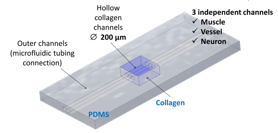
Scheme of the microfluidic chip
Figure 1 : Scheme of the microfluidic chip

To induce cellular self-organization, Institut Curie first textured the collagen I to create hollow cylindric channels of 200µm in diameter spaced 200µm apart inside the collagen bulk. Muscle cells are then seeded inside. This allows the cells to adhere, proliferate, grow and organize into myofibers. Besides the complexity of the chip architecture in structuring the scaffold, another challenge is in the seeding itself. In typical chips made out of polymers, cells are seeded manually with a pipette. With hydrogels, however, the seeding should be finely controlled. Hydrogels are highly fragile and the non-controlled pipetting on cells leads to hydrogel distortions and even to hydrogel detachment from the chip.

Movie and image courtesy of Manh Louis Nguyen, MMBM team , Institut Curie

To overcome this issue, a Flow-EZ pressure controller was used to gently inject cells inside the collagen channels. The pulse free injection preserves the architecture of the 3D hydrogel and promotes improved cell survival when compared to the acute shear stress associated with manual pipetting. The difference between these two modes of injection are illustrated in the videos below:

For more information or a technical discussion

Contact us
Logo fluigent green and blue

67 avenue de Fontainebleau
94 270 Le Kremlin-Bicêtre

Research

  • Microfluidic Research Applications
  • Instruments
  • Software solutions
  • Packages

Industrial

  • Products

Resources

  • Microfluidic Application Notes
  • Microfluidics case studies
  • Expert Reviews: Basics of Microfluidics
  • Interviews & Testimonials

Support & Tools

  • Documentation
  • Download software

Company

  • About us
  • Team
  • Events & Webinars
  • Newsletter
  • Fluigent’s Distributors
  • Careers

Legal

  • Terms & Conditions of Sale
  • Legal Terms & Privacy Policy