Microfluidic Immunofluorescence: Improving Staining Reproducibility for Microscopy
This page covers immunofluorescence and immunolabeling workflows run inside microfluidic chips and imaging chambers, where an automated system delivers each reagent under flow control.
Key takeaways
- Microfluidic immunofluorescence runs the fixation-to-imaging staining sequence inside a chip or closed imaging chamber. Each reagent is delivered by programmed flow rather than by hand pipetting [1].
- In a published application note, an automated immunofluorescence protocol ran in 4 h 30 min. The manual equivalent can take up to 1 h 30 min longer [1].
- Automated IBEX multiplex imaging alternates antibody labeling, imaging and fluorophore bleaching. A six-cycle, 25-plex experiment typically runs in about 12 hours [2].
- A single instrument can deliver up to 10 solutions under user-defined protocols and trigger microscope acquisition through TTL or TCP signals between steps [2,3].
- With the Aria perfusion system, variability between experiments drops to 0.5%, compared with 5.1% intra-operator and 8.1% inter-operator variability when pipetting by hand [3].
What is microfluidic immunofluorescence?
Microfluidic immunofluorescence is immunofluorescence staining performed inside a microfluidic chip or closed imaging chamber. Fixation, permeabilization, blocking, antibody and wash solutions are delivered in sequence by a flow control system. The sample stays in place while the fluids change around it, so each step runs at a programmed volume, flow rate and incubation time [1].
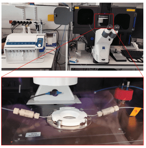

Figure 1. The Aria automated perfusion system delivers up to 10 solutions in sequence to a chip or imaging chamber [4].
Immunofluorescence is the most frequently applied technique for detecting and visualizing molecules in biological samples [5]. Its result depends on a multi-step protocol in which every step is a fluid exchange.
Moving that protocol from a bulk format into a microfluidic format reduces reagent and sample volumes and shortens incubation times. Automating the sequential steps also improves the reproducibility of the assay [4,6].
In practice, an automated system draws each solution from its own reservoir and routes it through a valve to the chip or chamber. It then holds or flushes the solution according to a saved protocol [4].
This page treats immunolabeling as a fluid-delivery problem. The antibodies are inputs to the workflow, and the flow control is the method.
Why manual immunofluorescence staining limits reproducibility
Manual immunofluorescence staining limits reproducibility because every fluid exchange depends on the operator. Pipetted volumes and flow rates vary from hand to hand, incubation times drift, and washes are applied unevenly. Multi-round experiments repeat each of these errors at every exchange, and each exchange consumes more reagent [1,3,7].
Pipetting variability. Manual pipetting is prone to inaccuracies in both solution volume and flow rate [1]. Fluigent measured 5.1% intra-operator and 8.1% inter-operator variability with a pipette, against 0.5% between experiments with automated delivery [3].
Abrupt injections. A pipette can deliver up to 1 mL in a few seconds through a conical tip, and successive injections differ from one another. Automated injections are smooth, identical and laminar [3].
Incubation-timing drift. Antibody incubation times and fluid exchange are among the technical challenges that a recent review of immunofluorescence optimization identifies [7].
Inconsistent washing under the microscope. Changing solutions by hand on the stage can displace the dish and its recorded regions of interest. It also risks losing the sample, contaminating it, spilling liquid over the microscope or running solutions in the wrong order [3].
Reagent consumption across rounds. Multi-solution experiments can extend to 4–5 days and require the scientist to be present throughout [3]. The original IBEX method was limited by manual execution, which restricted its throughput, speed and accessibility [2].
How microfluidic perfusion automates the staining workflow
Automation replaces each pipetting step with a programmed injection. The system selects a reservoir, delivers a set volume at a set flow rate, waits for the incubation time, flushes the line and moves to the next solution. The whole fixation-to-imaging sequence becomes one saved protocol that runs without the operator [1,3].
This approach builds on microfluidic perfusion, the controlled delivery of fluid through microscale channels and chambers [8].
In a perfused chamber, convective flow is the main factor that sets how a reagent reaches the cells. Flow rate and channel geometry therefore control the concentration and timing each cell experiences [8,9].
For each step of the protocol, the operator sets the flow rate, the volume dispensed and the incubation time in software. Protocols can be saved, recalled and edited, and the software reports the minimum volume required in each reservoir [3].

Figure 2. An immunolabeling protocol created in the Aria software: each step sets a reservoir, a flow rate, a volume or duration, and an incubation time [1].
Delivery timing. A solution leaving the valve reaches the sample only after it has filled the tubing between the two:
tarr = Vt / Q
Here tarr is the arrival time at the chip (min), Vt is the total internal and external tubing volume (µL), and Q is the flow rate (µL/min). The relation assumes a fully primed, bubble-free line.
Fluigent’s own example uses a setup with 300 µL of total tubing volume [3]. At 100 µL/min, a new solution therefore reaches the chamber about 3 minutes after the valve switches. This is why the tubing volume is calibrated, why the system prefills its lines with the successive solutions in order, and why the software calculates when each solution enters the flow cell [3].
Step volume. Each injection can be defined by volume or by duration, and the two are linked by V = Q × t. In Fluigent’s example, a step at 100 µL/min for 1 min delivers 100 µL [3].
A dedicated flush function clears the tubing with buffer between solutions to prevent carry-over. Preloading the system requires an additional 36 µL of liquid [3].
The immunofluorescence staining workflow, step by step
A standard immunofluorescence protocol runs through six fluid-handling stages: fixation, permeabilization, blocking, antibody incubation, washing and imaging [1]. Done by hand, each stage has its own source of variation. Under automated sequential reagent delivery, each stage becomes a programmed step with a fixed volume, flow rate and duration [1].
| Step | What varies when pipetted by hand | What automated delivery changes |
|---|---|---|
| Fixation | Exposure time and the force of each manual exchange around the sample | Runs as a programmed step, or before connection: one neuronal protocol fixed cells outside the system to keep fixative out of the internal fluidics [1,10] |
| Permeabilization | Volume added and the time before removal | Volume and flow rate set per step; a timed “wait” step fixes the incubation [1] |
| Blocking | Coverage of the whole chamber and incubation length | Controlled injection fills the chamber; the software times the incubation [1] |
| Antibody incubation | Incubation length and whether the dispensed volume reaches the whole sample | Set volume at a set flow rate, then a timed wait; automated IBEX labeled for 1 h at 37 °C on a heated stage [2] |
| Washing | Number, volume and force of rinses; abrupt pipette injections [3] | Wash defined by flow rate and volume or duration; tubing flushed between solutions [3] |
| Imaging | Handling the dish risks losing recorded regions of interest [3] | Sample stays on the stage; TTL or TCP signals launch acquisition and resume the protocol [2,3] |
Once the post-fixation sequence is automated, the operator is no longer the bottleneck. In the neuronal application note, the protocol allowed up to 12 microfluidic chips to be stained in a day with minimal user intervention [10].
Applications of microfluidic immunofluorescence
Microfluidic immunofluorescence applies wherever a staining or labeling protocol is a sequence of fluid exchanges. This covers fixed-cell and tissue staining, multiplex imaging and IBEX immunolabeling, FISH and DNA-PAINT labeling, and live-cell perfusion during imaging. In each family the instrument’s role is the same: it delivers the right solution at the right time without moving the sample [2,3].
Fixed-cell and tissue staining
Fixed samples are the most direct application. Permeabilization, blocking, antibody delivery and washing run as one automated sequence on a sample that stays in its chip or chamber. The protocol applies to cells on coverslips as well as to cells or tissues in microfluidic chips [1].
In the automated immunofluorescence application note, Aria stained primary human endothelial cells in an imaging chamber mounted on a widefield microscope. The full protocol took 4 h 30 min [1].
The neuronal cell immunofluorescence note automated immunolabeling of rat progenitor neurons across four microfluidic chips in one run, with up to three antibodies used simultaneously. The authors report clean staining with axons and dendrites preserved [10].
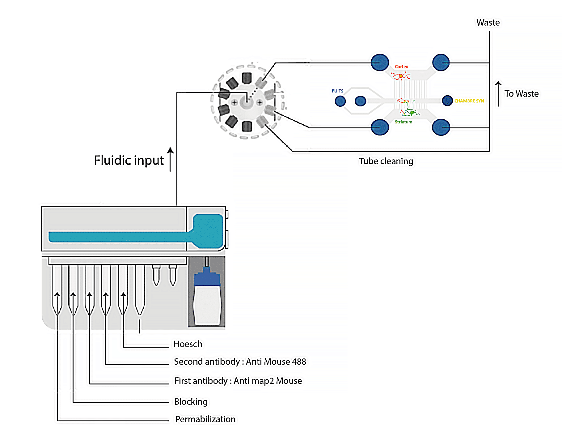

Figure 3. Immunofluorescence of neuron cells stained with anti-MAP2 for Microtubule-associated protein 2 (green) and with Hoechst for nuclei (blue). Images were acquired on a Nikon confocal microscope at 10X magnification [10].
Multiplex imaging and IBEX immunolabeling
Multiplex imaging extends staining into repeated rounds. Each round labels a panel, images it and inactivates the fluorophores before the next panel is applied to the same section. Because every round is a fluid sequence, the whole experiment can run as one programmed protocol rather than days of manual exchanges [2,11].
IBEX (iterative bleaching extends multiplexity) has been reported to reach more than 65 parameters in diverse tissues and is compatible with more than 250 commercially available antibodies [11]. It belongs to a wider family of cyclic immunofluorescence methods built on iterative staining and imaging [12].
The automated immunolabeling case study coupled Aria to a Leica THUNDER widefield microscope through TTL signals. The setup achieved more than 30 distinct antibody markers on the same tissue section, and was used to profile human lymph node, small intestine and skin [2].


Figure 4. (a) Schematic overview of automated IBEX protocol. (b) Images from human skin, five cycles, 15 of 19 parameters shown. Scale bars 200 µm (left), 25 µm (insets). Keratin 10 (K10), Keratin 14 (K14). Adapted from Radtke, Chu et al. (2022), as reproduced in the Fluigent case study [2,11].
FISH, DNA-PAINT and advanced labeling workflows
Sequential FISH and DNA-PAINT are labeling methods built on solution exchange, so each is a reagent-delivery sequence that an automated system can execute. seqFISH barcodes transcripts through successive rounds of hybridization, imaging and probe stripping [13]. Exchange-PAINT images multiple targets in sequence by exchanging the imager strands in solution [14].
Exchange-PAINT uses only a single dye and a single laser source. The underlying DNA-PAINT method achieved sub-10-nm resolution in vitro on synthetic DNA structures [14]. Its multiplexing therefore depends on accurate, repeatable exchange of solutions between imaging rounds.
Live-cell perfusion and imaging
Live-cell workflows apply the same sequential delivery to samples that are imaged while the fluids change. Instead of fixing and staining, the protocol perfuses defined solutions during acquisition, so the sample’s response to each exchange is recorded under flow control [15].
In a cardiac organ-on-a-chip protocol published in Nature Protocols in 2025, Aria delivered Tyrode’s solutions from 0.125 to 2.0 mM Ca²⁺ to microtissues on a stage heated to 37 °C [15,16]. Videos were acquired during the final 20 seconds of each perfusion step [15].
The live-cell imaging under perfusion case study reports that calcium transient amplitude increased with rising extracellular calcium [15].

Figure 5. Aria integrated with a cardiac organ-on-a-chip on a heated microscope stage, adapted from the supplementary material of the original protocol [15,16].
Imaging compatibility: fluorescence and confocal microscopy
Microfluidic immunofluorescence works with standard fluorescence and confocal microscopes because the chip or chamber stays on the microscope stage throughout. Staining and acquisition alternate without unmounting the sample, so the field of view and recorded regions of interest are kept from one round to the next [2,3].
The perfusion system and the microscope communicate through TTL or TCP signals. A step can send a signal when it starts, when it ends, or both. A “wait for signal” function holds the protocol until the microscope reports that acquisition is complete, and proceeds automatically after 12 hours if no signal arrives [3].
Published demonstrations cover several imaging platforms. These include widefield imaging on a Zeiss Axio Observer [1] and a Leica THUNDER [2], and spinning-disk confocal imaging of neuronal chips, which were disconnected after staining [10]. Synchronization with Zeiss ZEN software is possible over TCP/IP [15].
Registration across rounds is handled in image processing. In automated IBEX, a Hoechst nuclear label served as the fiducial and the images were aligned with the open-source SimpleITK software [2,11]. The IBEX protocol itself is designed for slides and nonproprietary imaging chambers so it can be adopted across imaging platforms [11].
Tools for automated immunofluorescence staining
Fluigent supplies the instrumentation for microfluidic immunofluorescence: an integrated instrument for sequential reagent delivery, a modular pack for laboratories that assemble their own setup, and chips designed for microscopy. Each runs the staining protocol under flow control, and the right choice depends on how integrated the setup needs to be.
- Aria automated perfusion system delivers up to 10 solutions in sequence into a chamber or microfluidic chip under user-defined protocols. Flow rates range from 3.2 µL/min to 1 mL/min for water, depending on the flow sensor fitted [3].
- Immunostaining Pack is a ready-to-use sequential-injection bundle that delivers up to 10 fluids to a single cell culture chamber. It combines a pressure-based flow controller, a flow sensor, a 10-position valve and automation software [4].
- Cell culture and microscopy chips are the substrates the staining workflow runs in. The range includes the live-cell imaging chamber used in the automated immunofluorescence application note [1].
Frequently Asked Questions
What formats can automated staining run on: chips, chamber slides or well plates?
Automated staining runs on closed formats with an inlet and an outlet: microfluidic chips, flow chambers, and imaging chambers holding coverslips or tissue sections. The published Aria workflows used a coverslip imaging chamber, PDMS chips and a closed bath tissue chamber. The single-output configuration delivers up to 10 fluids into one sample. The serial-output configuration, connected to a multi-position valve, delivers up to 10 fluids into 9 samples.
What flow rate should antibody incubation and washing steps use?
There is no single correct value. Flow rate is set for each step of the protocol, within the instrument’s range of 3.2–80 µL/min or 40 µL/min–1 mL/min for water, depending on the flow sensor fitted. Fluigent’s documentation uses 100 µL/min as its example for injection and tubing flush, and incubations then run as timed wait steps. Fragile samples need gentle flow. In microfluidic chambers, fluidic stresses can tear cells or detach them from their substrate, and the shear a sample experiences depends on both channel geometry and flow rate.
How many staining and stripping rounds can a multiplex experiment run?
The number of rounds is set by the antibody panel rather than by the fluidics. Automated IBEX repeats labeling, imaging and bleaching until the target number of parameters is reached. Reported automated runs include a six-cycle, 25-plex experiment in about 12 hours and five-cycle, 19-parameter imaging of human skin. IBEX inactivates fluorophores by chemical bleaching rather than by stripping antibodies. The method has been reported to enable imaging of more than 65 parameters in diverse tissues.
Related Expertises
-
Expert Reviews: Basics of Microfluidics Optimizing Microfluidic Perfusion: Best Practices and Innovations Read more
-
Microfluidic Application Notes Automating calcium imaging in neural cells with Fluigent’s Aria Read more
-
Microfluidic Application Notes Automating Neuronal Cell Immunofluorescence in Microfluidic Chips Read more
-
Microfluidics Case Studies University of Rochester: A tissue chip platform for real-time sensing of secreted inflammatory markers using ARIA Read more
-
Microfluidic Application Notes Automated Immunofluorescence using Aria Read more
-
Microfluidics Case Studies Automated immunolabeling to perform highly multiplexed tissue imaging with ARIA Read more
Sources
- Fluigent. (2023). Automated immunofluorescence using Aria [Application note]. https://www.fluigent.com/resources-support/expertise/application-notes/automated-immunofluorescence/
- Fluigent. (2022). Automated immunolabeling to perform highly multiplexed tissue imaging with ARIA [Case study]. https://www.fluigent.com/resources-support/expertise/customer-case-studies/automated-immunolabeling/
- Fluigent. (2022). Aria, an automated perfusion system. https://www.fluigent.com/research/instruments/aria/
- Fluigent. (2022). Immunostaining Pack. https://www.fluigent.com/research/instruments/packages/application-packages/immunostaining_package/
- Zaqout, S., Becker, L.-L., & Kaindl, A. M. (2020). Immunofluorescence staining of paraffin sections step by step. Frontiers in Neuroanatomy, 14, 582218. https://doi.org/10.3389/fnana.2020.582218
- Ciftlik, A. T., Lehr, H.-A., & Gijs, M. A. M. (2013). Microfluidic processor allows rapid HER2 immunohistochemistry of breast carcinomas and significantly reduces ambiguous (2+) read-outs. Proceedings of the National Academy of Sciences of the United States of America, 110(14), 5363–5368. https://doi.org/10.1073/pnas.1211273110
- Piña, R., Santos-Díaz, A. I., Orta-Salazar, E., Aguilar-Vazquez, A. R., Mantellero, C. A., Acosta-Galeana, I., Estrada-Mondragon, A., Prior-Gonzalez, M., Martinez-Cruz, J. I., & Rosas-Arellano, A. (2022). Ten approaches that improve immunostaining: A review of the latest advances for the optimization of immunofluorescence. International Journal of Molecular Sciences, 23(3), 1426. https://doi.org/10.3390/ijms23031426
- Fluigent. (2025). Optimizing microfluidic perfusion: Best practices and innovations. https://www.fluigent.com/resources-support/expertise/expertise-reviews/microfluidic-cell-biology/microfluidic-perfusion-innovation/
- Huber, D., Oskooei, A., Casadevall i Solvas, X., deMello, A., & Kaigala, G. V. (2018). Hydrodynamics in cell studies. Chemical Reviews, 118(4), 2042–2079. https://doi.org/10.1021/acs.chemrev.7b00317
- Fluigent. (2023). Automating neuronal cell immunofluorescence in microfluidic chips [Application note]. https://www.fluigent.com/resources-support/expertise/application-notes/neuronal-cell-immunofluorescence/
- Radtke, A. J., Chu, C. J., Yaniv, Z., Yao, L., Marr, J., Beuschel, R. T., Ichise, H., Gola, A., Kabat, J., Lowekamp, B., Speranza, E., Croteau, J., Thakur, N., Jonigk, D., Davis, J. L., Hernandez, J. M., & Germain, R. N. (2022). IBEX: An iterative immunolabeling and chemical bleaching method for high-content imaging of diverse tissues. Nature Protocols, 17(2), 378–401. https://doi.org/10.1038/s41596-021-00644-9
- Lin, J.-R., Fallahi-Sichani, M., & Sorger, P. K. (2015). Highly multiplexed imaging of single cells using a high-throughput cyclic immunofluorescence method. Nature Communications, 6, 8390. https://doi.org/10.1038/ncomms9390
- Lubeck, E., Coskun, A. F., Zhiyentayev, T., Ahmad, M., & Cai, L. (2014). Single-cell in situ RNA profiling by sequential hybridization. Nature Methods, 11(4), 360–361. https://doi.org/10.1038/nmeth.2892
- Jungmann, R., Avendaño, M. S., Woehrstein, J. B., Dai, M., Shih, W. M., & Yin, P. (2014). Multiplexed 3D cellular super-resolution imaging with DNA-PAINT and Exchange-PAINT. Nature Methods, 11(3), 313–318. https://doi.org/10.1038/nmeth.2835
- Fluigent. (2025). Multi-parametric functional assays of cardiac organ-on-a-chip using live-cell microscopy and Fluigent, Aria [Case study]. https://www.fluigent.com/resources-support/expertise/customer-case-studies/calcium-transient-imaging-with-aria/
- Garcia, M. I., Dame, K., Charwat, V., Siemons, B. A., Finsberg, H., Bhardwaj, B., Yokosawa, R., Goswami, I., Bruckner, D., Wall, S., Ford, K. A., Healy, K., & Ribeiro, A. J. (2025). Human induced pluripotent stem cell-derived cardiomyocytes and their use in a cardiac organ-on-a-chip to assay electrophysiology, calcium and contractility. Nature Protocols. https://doi.org/10.1038/s41596-025-01166-4


