Webinar - Microfluidics Automation for High-Resolution Microscopy
Our webinar exploring the microfluidics automation for multiplexing high-resolution microscopy workflows.
Watch this video replay of the webinar and learn how on-stage staining can be streamlined using the Aria perfusion system, seamlessly integrated with ZEISS ZEN software, featuring the case study of single-molecule localization microscopy techniques like DNA-PAINT and Exchange-PAINT.
Speakers:
Anel Rakhmatullina: Life-Science Applications Engineer, Fluigent
Dr.Philipp Seidel: Product Manager, Business Sector Life Science, ZEISS Research Microscopy Solution (RMS)
Agenda:
✅ Introduction to On-Stage Perfusion Optimization for Microscopy Applications
Overview of use of Aria with flow cells and microfluidic chips for immunolabelling applications, FISH, and DNA-PAINT
Presented by Anel, Fluigent
✅ Integration of Aria with ZEISS ZEN Microscopy Software
Next, learn how Aria integrates directly with the ZEISS ZEN Microscopy Software, enabling automated, precise control during imaging workflows.
Presented by Dr. Philipp Seidel, ZEISS RMS
✅ Case Study: DNA-PAINT for Single-Molecule Localization Microscopy
Then, dive into a real-world case study showcasing the use of DNA-PAINT for super-resolution imaging at the single-molecule level.
Presented by Dr. Philipp Seidel, ZEISS RMS
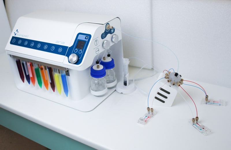
Explore the Versatility of Aria: The Automated Perfusion System
As a researcher performing live-cell imaging & labeling experiments, you know how they can be expensive (reagents, sample, microscope), time-consuming and stressful. A mistaken operation can ruin the whole experiment. Helping researchers to avoid those risks, to gain productivity, and to get the best of each experiment, Fluigent has created Aria.
Aria’s advanced automated perfusion system is designed to support a wide range of experimental workflows, including those integrated with microscopy. Whether you’re conducting live-cell imaging or complex staining protocols, Aria streamlines your processes for greater efficiency and precision.
Explore how the Chu Lab is advancing ocular research
Based at the UCL Institute of Ophthalmology in partnership with Moorfields Eye Hospital, the Chu Lab—led by Dr. Colin Chu—focuses on ocular immunology, spatial biology, immune cell imaging, and gene therapy.
In this case study, discover how they overcame the limitations of single-cell genomics by using automated immunolabeling for multiplex optical imaging—enabling deep phenotyping and spatial analysis of cells in complex tissues.
Discover how Aria can transform your research through examples of applications:
- Neuronal Cell Immunolabeling in Microfluidic Chips : Aria enables parallel immunofluorescence labeling of neuronal cells across up to four microfluidic chips simultaneously. By automating the staining process, it significantly reduces hands-on time while ensuring consistent, high-quality results—minimizing cell stress and eliminating antibody residue.
- Calcium Imaging in Neurons: With Aria, calcium imaging experiments are fully automated, allowing for faster, more reliable data acquisition. The system ensures stable and reproducible recordings, making it ideal for high-throughput microfluidic studies.
- Cancer Cell Capture and Labeling : Aria supports the complete automation of capturing and labeling MDA-MB-231 breast cancer cells using up to 10 different reagents. From surface treatment and bead injection to cell suspension and immunostaining, Aria simplifies complex workflows—reducing processing time, cutting reagent consumption, and minimizing human error.
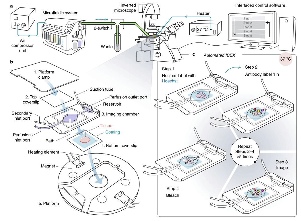
WEBINAR Replay – Automated IBEX multiplex immunohistochemistry using Fluigent’s ARIA
Ready to automate your immunohistochemistry workflows?
Access our insightful webinar and join Dr. Colin Chu from the UCL Institute of Ophthalmology (UK) as he presents a powerful technique developed with the Germain Research Group at the US NIH: Iterative Bleaching Extends Multiplexity.
Using Fluigent’s Aria automated perfusion solution, the team successfully labeled up to 40 proteins in a single tissue section—pushing the boundaries of multiplex imaging with microfluidics.
Discover the method and its impact.