Why is it important to control shear stress in your microfluidic experiments?

Mechanical forces are potent regulators of cellular structures and functions in both health and disease. As a result of their unique location, endothelial cells experience several mechanical forces: pressure, created by the hydrostatic forces of blood within the blood vessel; circumferential stretch generated by cardiac pulsation; and shear stress, the dragging frictional force created by blood flow. Of these forces, shear stress is particularly important as it stimulates the release of vasoactive substances and changes gene expression, cell metabolism, and cell morphology.

Physical characteristics of fluid shear stress

The shear stress τ is defined as the ratio of the tangential force to the surface area to which it is applied, and is expressed as a pressure. Fluid shear stress correlates with fluid velocity and with viscosity. For a Newtonian fluid, for which the viscosity is constant, the shear stress depends on the viscosity of the fluid and the shear rate. Therefore, for Newtonian fluids it may be computed according to Newton’s law with the following relation:

A flowing fluid displays a parabolic velocity profile (fluid velocity is fastest at the center than near the channel wall).

However, the blood in a non-Newtonian fluid and its viscosity is not constant. An apparent viscosity can be attributed, i.e., the viscosity of a Newtonian fluid showing the same relationship between the flow rate Q and the pressure differential ΔP, according to the Poiseuille’s law.

Use our Shear Stress Calculator to determine your experimental parameters. Define the flow rate or the pressure to be applied in your system, the dimension of microfluidic chip and tubing length in order to apply the correct shear stress for your cell culture under flow conditions.

laminar flow inside a circular channel
Figure 1: Representation of the flow velocity profile (left) and shear rate distribution (right) of a laminar flow inside a circular channel

In vivo fluid shear stress

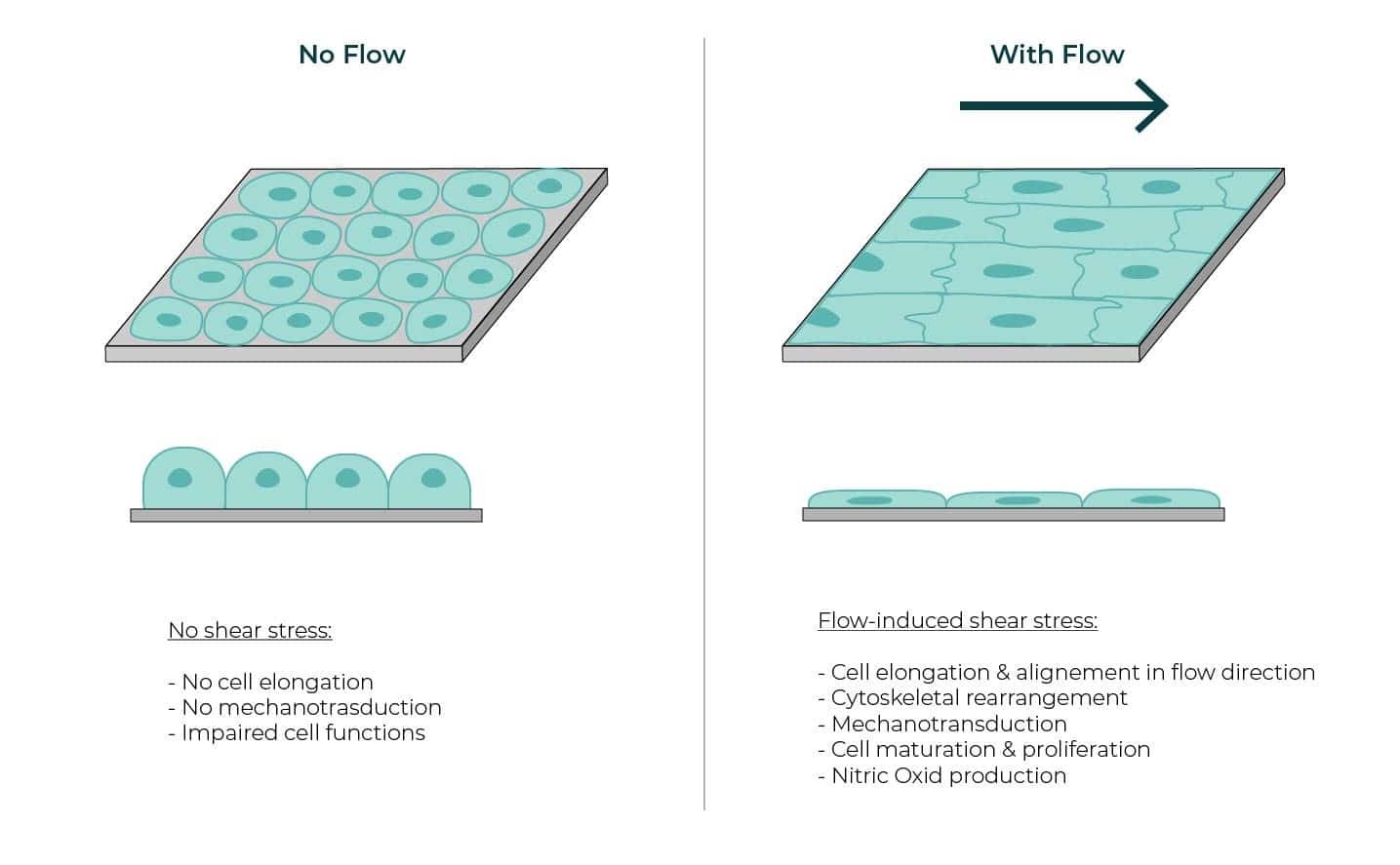
Cells are submitted to a variety of mechanical forces on a daily basis. Endothelial cells (ECs) lining blood vessels are constantly exposed to shear stress resulting from the blood flow. Shear stress corresponds to the dragging force generated by the friction between a moving fluid and the cells forming the inner lining of a vessel. Other cells exposed to shear stress include kidney epithelial cells or endothelial cells forming the lymphatic vessels1. This influences their behavior and gene expression. Cells exposed to a rapid and unidirectional blood flow display a spindle-like shape and align their longitudinal axis parallel to the direction of blood flow2 (Figure 2). This is caused by cytoskeletal remodeling to minimize strain generated by the shear stress3-4.

These mechanical forces cause morphological changes and trigger biochemical and biological events. Cells can sense mechanical cues (though their membrane receptors, ion channels, cytoskeleton and nuclear envelop proteins) and transduce them into molecular signaling cascades, a mechanism known as mechanotransduction. ECs respond to physical forces such as shear stress to maintain vascular homeostasis.

flow-induced shear stress
Figure 2: Cells sense and react to flow-induced shear stress

Several investigations have revealed that shear stress regulates major EC functions including angiogenesis, vessel remodeling, and cell fate5. Acute changes in stretch and shear stress induce transient changes including the release of vasoactive agent and alterations in vessel diameter. Thus, physiologic stresses and strains exert vasoprotective roles via the production of NO (Nitric Oxid) by the endothelium. NO stimulates the relaxation of vascular smooth muscle and regulates vascular resistance and blood pressure6. However, disrupted shear stress can drive abnormal endothelial cell behavior and vascular wall remodeling, causing inflammation that can be associated with cardiovascular disorders, including hypertension, atherosclerosis and stroke.

In vivo, the wall shear stress is ~1-2 Pa (10-20 dyne/cm2) in arteries, ~5 Pa in small arterioles7, ~2 Pa in venules, and ~0.1 Pa (dyne/cm2) in the vena cava7-10. A detailed list of in vivo shear stress values can be found in Table 1.

In vivo shear stress values

Physiological shear stress values range from 0.1 to more than 100 dyne/cm² and depend on the tissue (e.g., kidney, lung, heart) or the vessel type for blood vessels (e.g., artery or vein). It also differs from one organism to another (e.g., mouse versus human). The shear stress is often measured in dyne/cm² (dyn/cm²) or in Pascal (Pa). Of note, 1 Pa = 10 dyn/cm2

Cell typesShear stress value (Pa)Shear stress value (dyn/cm2)
Arteries111-2 Pa10-20 dyn/cm2
Human common femoral artery120.3-0.5 Pa3-5 dyn/cm2
Carotid artery121.1-1.3 Pa11-13 dyn/cm2
Veins110.1-0.6 Pa1-6 dyn/cm2
Venules112-4 Pa20-40 dyn/cm2
Arterioles116-8 Pa60-80 dyn/cm2
Capillaries110.5-2 Pa5-20 dyn/cm2
Glomerular capillaries130.1-9.5 Pa1-95 dyn/cm2
Embryonic lung140.01-0.1 Pa0.1-1 dyn/cm2
Airway epithelial cells150.05-0.3 Pa0.5-3 dyn/cm2
Alveolar epithelial cells160.4-1.5 Pa4-15 dyn/cm2
Mouse embryonic kidney170.04-0.5 Pa0.4-5 dyn/cm2
Human kidney180.03-0.12 Pa0.3-1.2 dyn/cm2
Cornelial epithelial cells190.4-0.8 Pa4-8 dyn/cm2
Osteocytes200.8-3 Pa8-30 dyn/cm2

Table 1: In vivo shear stress value of different types of blood vessels and other tissues

Diversity of flow-induced shear stress

Blood flow dynamics influence EC cytoskeletal organization and inflammatory state. Indeed, ECs are sensitive to small variations in magnitude, but also in the direction and regularity of blood flow-induced shear stress21. ECs experienced several types of shear stress:

luminal flow within the vessel lumen that is parallel to the cell and acts on EC apical side

– pulsation across the endothelium produced by heartbeats which rather impacts cell–cell junctions

– interstitial flow resulting from liquid flowing among the interstitial space and acting on EC basal side.

Luminal – Steady laminar flow

From a pure physical aspect, Reynolds number (for a tube , with  the fluid density,  the flow velocity, and  the tube diameter) helps to predict flow patterns in different fluid flow situations. At low Reynolds number (typically Re < 1000), the flow is laminar: fluid flows in parallel layers, with no disruption between them, as opposed to turbulent flow. As the dimensions are in micrometer scale in most tissues and organs (e. g., capillaries are about 8-10µm in diameter), Reynolds number is low (typically < 10). Flows are mainly laminar, with a characteristic velocity flow profile (Figure 1). Vascular flows are most typically uniaxial and laminar. In relatively straight vascular segments, flow streamlines are largely undisturbed and remain mostly parallel to the vascular wall22 (Figure 3A). Steady laminar shear stress is a determinant of normal vascular function through the orchestration of NO production and anti-platelets aggregation mechanism.

Luminal – Turbulent flow

Blood flow within vessels is generally laminar and streamlined. Abrupt changes in vessel geometry due to branching, sharp turns or stenosis can disturb the laminar blood flow, causing secondary flows in the form of vortices (Figure 3B). ECs lining those turbulent segments or stagnant are much rounder in shape and do not have a uniform orientation. Flow disturbance often correlates with the localization of vascular diseases including atherosclerosis, aortic valve calcification, and inflammation and thrombosis.

Pulsatile flow

Pulsatile flow is mainly observed in large vessels such as arteries. It is a consequence of rhythmic heartbeat. At each beat, aortic flow reaches a peak (systole), then diminishes to a low level (diastole) until the next beat. This produces pulsatile flow instead of a continuous flow (Figure 3A). Though pulsatile, the flow remains laminar. The velocity flow profile varies as a function of time23. This has an impact on the shear stress as it is derived from flow velocity. Pulsatile flow is usually introduced in blood vessel-on-chip models to simulate the actual pulsatile blood flow in human circulation24.

Interstitial flow

Interstitial flow arises from fluid movement within the extracellular matrix surrounding the ECs, shearing them on their basal side25 (Figure 3C). Fluid flow carries large proteins through the interstitial space and mechanically stimulates ECs. Several studies have demonstrated that shear flow induced by interstitial fluid was crucial for physiological cell responses such as cell differentiation26-27. Interstitial fluid flows at a lower velocity as compared to blood flow within vessels because of high resistance exerted by the extracellular matrix. Flow velocity profile and subsequent shear flow is also more difficult to define due to the complex architecture of the extracellular matrix and as the fluid moves around the cell-matrix interface in all directions. Interstitial flow significantly enhances vascular sprout formation, network extension, and the development of branching networks in a magnitude-dependent manner28.

distribution of flow velocities
Figure 3: The different types of flow (Arrows represent the distribution of flow velocities)

Studying the effect of shear stress in vitro in relevant physiological systems

Traditionally, endothelial cells have been grown and studied in the absence of flow and mechanical stimulations on stiff substrates such as plastic or glass. These cells are not subjected to the physical forces that endothelial cells endure in vivo, thus the results of these experiments often do not mimic those observed in the body. The field of vascular biology now realizes that an intricate analysis of endothelial signaling mechanisms requires complex in vitro systems to mimic in vivo conditions.

In vitro platforms to apply defined mechanical stimuli to cells are critical to understanding how strains and shear stresses affect endothelial and other cells. Early studies have been performed using macro scale, custom built devices reviewed here29. For the past 10 years, the development of microfabrication and bioengineering approaches has given rise to microfluidic devices that offer the ability to control or alter the cell’s physical environment.

These devices have been used to grow ECs under dynamic flow conditions to impose shear stress (by direct control of the fluid flow rate) and to analyze their responses at the cellular and molecular levels. Different systems have been developed to address specific aspects of how shear and strain affect EC functions. These systems have demonstrated that ECs alter their morphology, function, and gene expression in response to shear stress. In vitro models of the human microvasculature to study the effect of luminal flow on cancer cell migration30 have contributed to a better understanding of the metastatic cascade process (extravasation and subsequent interstitial migration). Luminal flow significantly promoted the extravasation potential of tumor cells as compared to static conditions, with an average intravascular speed of tumor cells of ~ 12.5 μm/h under flow, as compared to ~ 9.4 μm/h under static conditions. This reveals the important role of fluid flow during metastatic extravasation and invasion30. In another study, a microfluidic device was developed to gain insights into how interstitial flow affects breast cancer cell invasion31. By performing live cell imaging under well-controlled flow conditions, the authors found that compared to static flow conditions, interstitial flow increases the percentage of cells that become migratory and increased their average migration speed. Fluid flow within the tumor microenvironment can enhance tumor cell invasion by directing a subpopulation of tumor cells in the flow direction; i.e., towards the draining lymphatic vessels, a major route of metastasis31. These observations demonstrate how important it is to consider both laminar and interstitial flows in tumor models, as they affect tumor cell extravasation and invasion.

Implementing shear stress in in vitro culture systems

Choosing the best fluid delivery instrument

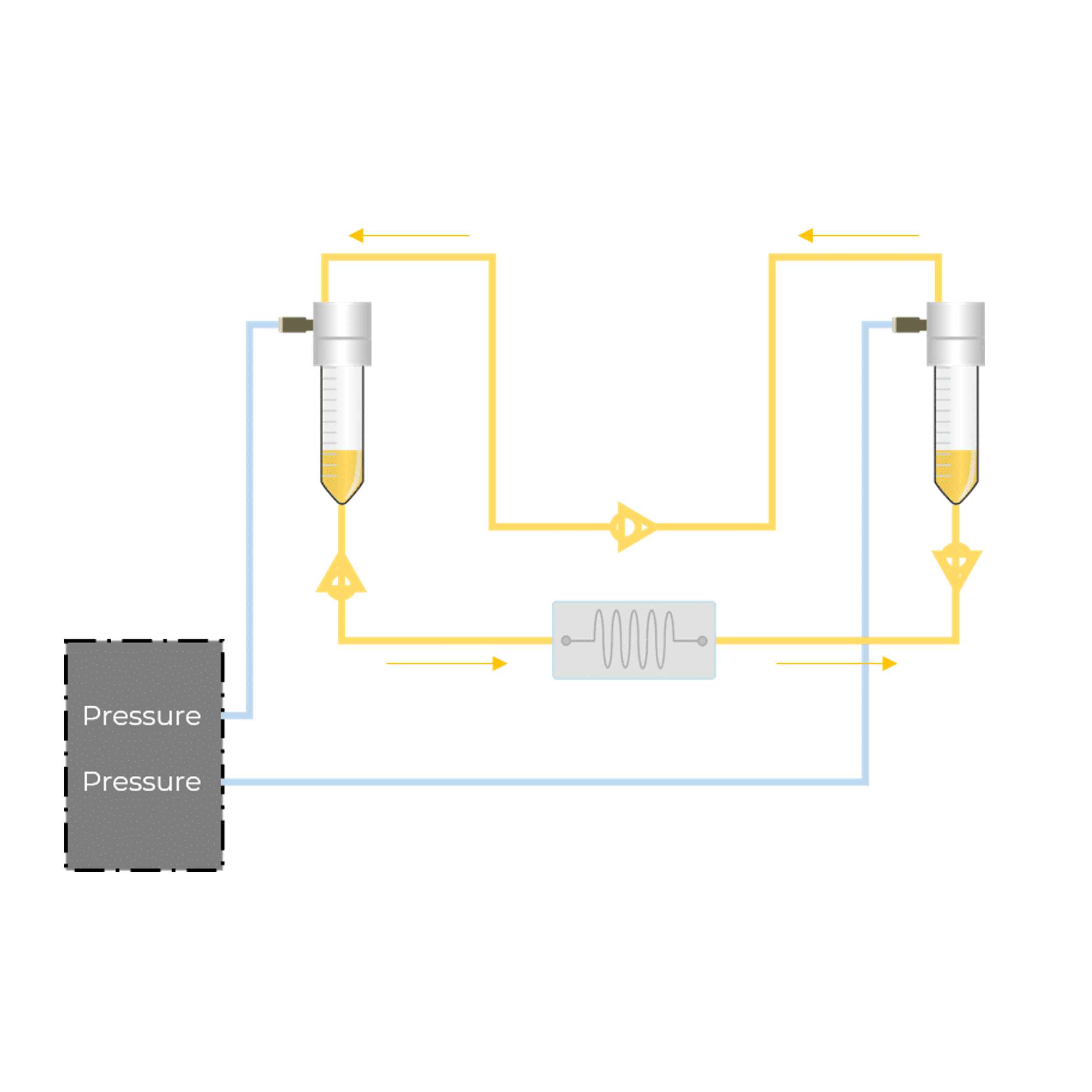
As described in this review, shear stress is a mechanical force that strongly influences cell morphology and behavior. Several microfluidic devices compatible with fluid perfusion and application of different shear stress strengths for the analysis of cellular functions now exist. To implement shear stress into microfluidic devices, it is essential to accurately control fluid delivery. The choice of flow control instrument is crucial. Available options include syringe pumps, peristaltic pumps or pressure-based flow controllers. A pressure-based system can provide well-controlled and steady flow rates as opposed to other devices such as peristaltic pumps. We have compared the impact of the perfusion system on endothelial cells seeded in microfluidic chips using either a peristaltic pump or pressure-based flow controllers.

This test highlights the importance of flow stability in vascular models. It is important to choose an instrument providing instantaneous response time and preventing flow rate oscillations.
Pressure controllers comply with these requirements as they offer the fastest response time and the most accurate flow control. We have compared the response time as well as the flow stability of high precision syringe pump and flow controllers.

Shear stress calculation in a microfluidic device

The shear stress applied on cells exposed to constant flow within a microfluidic chip can be determined using the continuity and Navier-Stokes equations. Following are the flow velocity profiles and shear stress equations for two common microfluidic channel geometries: circular and rectangular channels.

Circular channel  

For a circular cross-section of diameter d, the fully developed velocity profile in cylindrical coordinates (r,θ,z) follows the equation32:

Shear stress calculation in circular channel

With Q the flow rate, and r the radial distance from the centerline of the channel. The distribution of flow velocities follows a parabolic profile, and the maximum velocity is for r = 0, at the center of the channel.

It is of interest to determine the shear stress distribution at the channel wall (r = d/2), as this is where cells are located in the chip. To do so, the strain rate should be calculated by differentiating the flow velocity with respect to r. We get:

With µ the dynamic viscosity, Q the flow rate. This indicates that the shear stress remains constant along the channel walls and is a function of the viscosity, flow rate, and channel diameter. At constant channel diameter and viscosity, the higher the flow rate, the higher the shear stress.

Rectangular channel

In microfluidic devices with rectangular channels, the flow velocity profile and subsequent shear stress are more complex. Wall shear stress is not constant and varies across the top, bottom, and side walls of the channel. However, the geometry can be simplified by considering two infinite parallel plates instead of closed channels. Under this assumption, the shear stress follows the equation:

Shear stress calculation in rectangular channel

With Q the flow rate, η the dynamic viscosity, w, and h the channel width and height, respectively.

Use our Shear Stress Calculator to determine your experimental parameters. Define the flow rate or the pressure to be applied in your system, the dimension of microfluidic chip and tubing length in order to apply the correct shear stress for your cell culture under flow conditions.

References

  1. Ferrell N. et al. Application of physiological shear stress to renal tubular epithelial cells. Methods Cell Biol (2019). doi: 10.1016/bs.mcb.2019.04.010
  2. Dewey C.F Jr. et al. The dynamic response of vascular endothelial cells to fluid shear stress. J Biomech Eng (1981). doi: 10.1115/1.3138276.
  3. Malek A.M & Izumo S. Mechanism of endothelial cell shape change and cytoskeletal remodeling in response to fluid shear stress. J Cell Sci (1996). doi: 10.1242/jcs.109.4.713.
  4. Helmke B.P, Rosen A.B & Davies P.F. Mapping mechanical strain of an endogenous cytoskeletal network in living endothelial cells. Biophys J (2003). doi: 10.1016/S0006-3495(03)75074-7.
  5. Campinho P, Vilfan A, Vermot J. Blood flow forces in shaping the vascular system: a focus on endothelial cell behavior. Front Physiol (2020) doi: 10.3389/fphys.2020.00552).
  6. Davies P.F. Hemodynamic shear stress and the endothelium in cardiovascular pathophysiology. Nat Clin Pract Cardiovasc Med (2009). doi: 10.1038/ncpcardio1397.
  7. Papaioannou T & Stefanadis C. Vascular wall shear stress: basic principles and methods. Hell. J. Cardiol. (2005)
  8. Lipowsky, H. H., Kovalcheck, S. & Zweifach, B. W. The distribution of blood rheological parameters in the microvasculature of cat mesentery. Circ Res (1978). doi: 10.1161/01.res.43.5.738.
  9. Natarajan, M., et al. Hemodynamic flow-induced mechanotransduction signaling influences the radiation response of the vascular endothelium. Radiat Res (2016). doi: 10.1667/RR14410.1.
  10. Chatterjee S. Endothelial mechanotransduction, redox signaling and the regulation of vascular inflammatory pathways. Front Physiol (2018). doi: 10.3389/fphys.2018.00524.
  11. Lipowsky H.H., et al. The distribution of blood rheological parameters in the microvasculature of cat mesentery. Circ Res. (1978). doi: 10.1161/01.res.43.5.738.
  12. Reneman R.S., & Hoeks A.P.G. Wall shear stress as measured in vivo: consequences for the design of the arterial system. Med Biol Eng Comput (2008). doi: 10.1007/s11517-008-0330-2.
  13. Remuzzi A., et al. Three-dimensional reconstructed glomerular capillary network: Blood flow distribution and local filtration. Am J Physiol (1992). doi: 10.1152/ajprenal.1992.263.3.F562.
  14. Conrad L., et al. The biomechanical basis of biased epithelial tube elongation in lung and kidney development. Development (2021). doi: 10.1242/dev.194209.
  15. Sidhaye V.K., et al. Shear stress regulates aquaporin-5 and airway epithelial barrier function. PNAS (2008). doi: 10.1073/pnas.0712287105.
  16. Flitney E.W., et al. Insights into the mechanical properties of epithelial cells: the effects of shear stress on the assembly and remodeling of keratin intermediate filaments. FASEB J (2009). doi: 10.1096/fj.08-124453.
  17. Kimura H., et al. Effect of fluid shear stress on in vitro cultured ureteric bud cells. Biomicrofluidics (2018). doi: 10.1063/1.5035328.
  18. Ross E.J., et al. Three dimensional modeling of biologically relevant fluid shear stress in human renal tubule cells mimics in vivo transcriptional profiles. Sci Rep (2021). doi: 10.1038/s41598-021-93570-5.
  19. Molladavoodi S., et al. Corneal epithelial cells exposed to shear stress show altered cytoskeleton and migratory behaviour. Plos One (2017). doi: 10.1371/journal.pone.0178981.
  20. Weinbaum S., et al. A model for the excitation of osteocytes by mechanical loading-induced bone fluid shear stresses. J Biomech (1994). doi: 10.1016/0021-9290(94)90010-8.
  21. Givens, C., & Tzima, E. Endothelial mechanosignaling: does one sensor fit all? Antioxid Redox Signal (2016). doi: 10.1089/ars.2015.6493.
  22. Hahn, C. & Schwartz, M. A. Mechanotransduction in vascular physiology and atherogenesis. Nat Rev Mol Cell Biol (2009). doi: 10.1038/nrm2596.
  23. Zamir, M. Hemo-Dynamics. (2016). Book
  24. Liu, X. F et al. Biological factors in plasma from diabetes mellitus patients enhance hyperglycaemia and pulsatile shear stress-induced endothelial cell apoptosis. Integr Biol (Camb) (2014). doi: 10.1039/c3ib40265g.
  25. Yao, W., Li, Y. & Ding, G. Interstitial fluid flow: The mechanical environment of cells and foundation of meridians. Evid based Complement Alternat Med (2012). doi: 10.1155/2012/853516.
  26. Ng, C. P., Hinz, B. & Swartz, M. A. Interstitial fluid flow induces myofibroblast differentiation and collagen alignment in vitro. J Cell Sci (2005). doi: 10.1242/jcs.02605.
  27. Ng, C. P. & Swartz, M. A. Mechanisms of interstitial flow-induced remodeling of fibroblast-collagen cultures. Ann Biomed Eng (2006). doi: 10.1007/s10439-005-9067-3.
  28. Abe Y., et al. Balance of interstitial flow magnitude and vascular endothelial growth factor concentration modulates three-dimensional microvascular network formation. APL Bioeng (2019). doi: 10.1063/1.5094735.
  29. Brown T. D. Techniques for mechanical stimulation of cells in vitro: a review. Journal of Biomechanics (2000). doi: 10.1016/s0021-9290(99)00177-3.
  30. Hajal, C., et al. The effects of luminal and trans-endothelial fluid flows on the extravasation and tissue invasion of tumor cells in a 3D in vitro microvascular platform. Biomaterials (2021). doi: 10.1016/j.biomaterials.2020.120470.
  31. Haessler, U., et al. Migration dynamics of breast cancer cells in a tunable 3D interstitial flow chamber. Integr Biol (2012). doi: 10.1039/c1ib00128k.
  32. Huber, D., et al. Hydrodynamics in Cell Studies. Chem Rev (2018). doi: 10.1021/acs.chemrev.7b00317.

For more information or a technical discussion

Contact us