Resource / Application notes
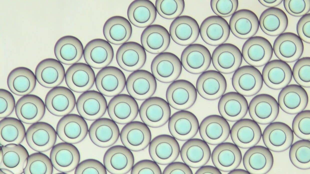
PLGA nanoparticle synthesis using 3D microfluidic hydrodynamic focusing
In this Application Note, PLGA nanoparticles with high monodispersity are generated using the Raydrop single emulsion developed and manufactured by Secoya, and Fluigent pressure-based flow controllers. The ability to synthesise PLGA nanoparticles in a more controllable and reproducible way creates possibilities to tailor surface properties and increase fields of application.