Mikrofluidik für Omics-Anwendungen
Was sind Omics?
„Omics“ bezieht sich auf Disziplinen, die sich mit der umfassenden Analyse biologischer Makromoleküle in einer Zelle, einem Gewebe oder einem Organismus befassen. Diese Disziplinen beinhalten die Analyse verschiedener biologischer Komponenten in großem Maßstab, typischerweise auf molekularer Ebene, um ein umfassendes Verständnis biologischer Systeme zu erlangen. Diese umfassende Analyse erfordert den Einsatz spezifischer Werkzeuge sowohl für biologische Experimente als auch für die Datenanalyse durch bioinformatische Ansätze.
Hier sind einige Schlüsseldisziplinen der Omics :
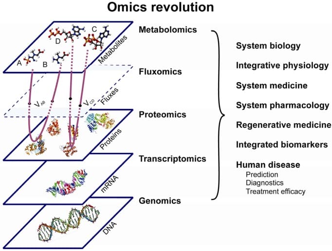
- Genomik: untersucht den gesamten DNA-Satz eines Organismus (Genom), um dessen Struktur, Funktion, Variationen und Wechselwirkungen zwischen den Genen zu verstehen.
- Transkriptomik: untersucht RNA-Moleküle innerhalb einer Zelle, einschließlich ihrer Arten, Häufigkeit und Veränderungen in den Genexpressionsmustern.
- Proteomik: untersucht die Gesamtheit der Proteine in einer Zelle, ihre Funktionen, Strukturen, Veränderungen, Wechselwirkungen und Häufigkeit.
- Metabolomik: analysiert die Gesamtheit der kleinen Moleküle oder Metaboliten, die am zellulären Stoffwechsel beteiligt sind, und liefert Erkenntnisse über Stoffwechselwege und physiologische Veränderungen.
- Epigenomik: erforscht Modifikationen und Veränderungen der Genexpression, die durch Faktoren außerhalb der DNA-Sequenz verursacht werden, wie DNA-Methylierung und Histon-Modifikationen.
- Metagenomik: konzentriert sich auf das genetische Material, das direkt aus Umweltproben gewonnen wird, und bietet Einblicke in mikrobielle Gemeinschaften und ihre genetische Vielfalt.

Abbildung 1: Die „Omics-Revolution“ – ein integrierter umfassender „Omics“-Ansatz, der Genomik, Transkriptomik, Proteomik, Metabolomik und Fluxomik zur Förderung der systemischen Wissenschaften und zur Diagnose und Behandlung menschlicher Krankheiten kombiniert (1).
Omics-Analysemethoden sind dabei, die Forschung in den Biowissenschaften radikal zu verändern. Ihre Fähigkeit, die Genome, Transkriptome oder Proteome einzelner Zellen zu bewerten, statt durchschnittliche Zustände innerhalb von Zellpopulationen zu beurteilen, stellt einen bedeutenden Sprung in verschiedenen Bereichen dar. So zum Beispiel in der Krebsbiologie, den Neurowissenschaften, der Therapie mit neuralen Stammzellen und anderen Gebieten.
Schlüsseltechniken der Omics-Disziplinen
Diese Omics-Disziplinen setzen fortschrittliche Technologien und Hochdurchsatztechniken ein, um große Datensätze zu erzeugen. Durch die Integration und Analyse dieser Datensätze können Forscher komplexe biologische Prozesse und Krankheitsmechanismen verstehen, Biomarker identifizieren und den Weg für eine personalisierte Medizin und gezielte Therapien ebnen.
Omics-Techniken sind weit verbreitet und variieren je nach dem spezifischen Omics-Bereich, der erforscht wird.
- PCR (Polymerase-Kettenreaktion): wird in der Genomik zur Vervielfältigung spezifischer DNA-Sequenzen verwendet und ermöglicht deren Analyse und Identifizierung.
- Next-Generation Sequencing (NGS): wird in der Genomik und Transkriptomik zur Sequenzierung von DNA und RNA verwendet und ermöglicht die Analyse von Genomen, Genexpression, Mutationen und Variationen in großem Maßstab.
- Massenspektrometrie (MS): wird in der Proteomik und Metabolomik eingesetzt, um Proteine oder Metaboliten in einer Probe zu identifizieren und zu quantifizieren, was Einblicke in ihre Strukturen, Veränderungen, Wechselwirkungen und Konzentrationen ermöglicht.
- Bildgebende Verfahren: werden in verschiedenen Bereichen eingesetzt, um molekulare Strukturen oder Verteilungen in Zellen oder Geweben sichtbar zu machen, zum Beispiel Fluoreszenzmikroskopie, Elektronenmikroskopie und bildgebende Massenspektrometrie.
- Microarrays: werden in der Genomik und Transkriptomik eingesetzt, um die Expressionsniveaus von Tausenden von Genen oder RNAs gleichzeitig zu analysieren, was ein Screening mit hohem Durchsatz und einen Vergleich der Genexpressionsmuster ermöglicht.
- Chromatographie: wird in der Metabolomik zur Trennung und Analyse komplexer Stoffwechselgemische auf der Grundlage ihrer chemischen Eigenschaften verwendet und hilft bei ihrer Identifizierung und Quantifizierung.
- Bioinformatik-Tools: Entscheidend für die Verarbeitung, Analyse und Interpretation der riesigen Datenmengen, die durch Omics-Techniken generiert werden, einschließlich rechnerischer Analyse, statistischer Modellierung und Datenintegration.

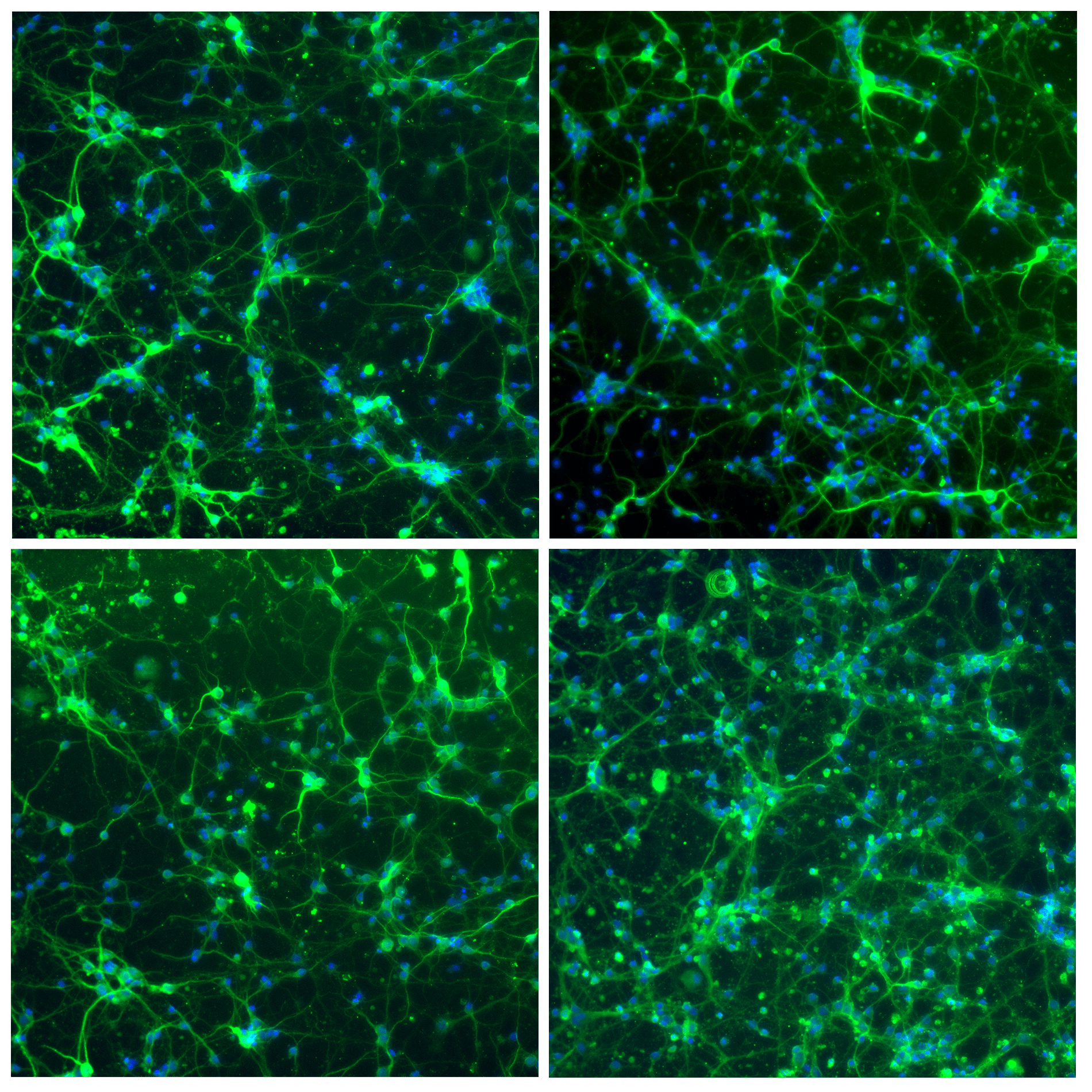
Abbildung 2: Immunfluoreszenz von Neuronenzellen, gefärbt für Mikrotubuli assoziiertes Protein 2 (grün) und für Zellkerne (blau). Die Bilder wurden mit einem konfokalen Mikroskop von Nikon bei 10-facher Vergrößerung aufgenommen.
Diese und andere Techniken entwickeln sich ständig weiter und verschmelzen mit fortschrittlichen Technologien, was zu einem umfassenden Verständnis biologischer Systeme beiträgt und Innovationen in den Biowissenschaften vorantreibt.
Präzise Flusshandhabung und der Beitrag von Fluigent für Omics
Die Revolution des Omics-Bereichs ist zu einem großen Teil den auf Mikrofluidik basierenden Techniken zu verdanken, bei denen die Zellen in Tröpfchen, Mikrokanäle oder Mikrozellen aufgeteilt werden, bevor sie der gewünschten Omics-Analyse unterzogen werden.
Die präzise Handhabung von Flüssen ist von grundlegender Bedeutung für verschiedene analytische Prozesse, die mit DNA, RNA, Proteinen und Metaboliten zu tun haben. Sie ist von entscheidender Bedeutung bei Schritten wie der Probenvorbereitung, bei der ein genaues Pipettieren, Verdünnen und Mischen der Proben erforderlich ist. Die Kombination von Automatisierung und präzisem Fluidhandeling erleichtert das Hochdurchsatz-Screening in der Genomik, Transkriptomik und Proteomik und ermöglicht die effiziente Verarbeitung großer Probenmengen.
Insgesamt ist die Gewährleistung von Genauigkeit und Reproduzierbarkeit bei der Probenvorbereitung, -trennung und -analyse entscheidend für die Generierung zuverlässiger Daten, die wesentlich zum Verständnis biologischer Systeme auf molekularer Ebene beitragen.
Fluigent hat sich zum Ziel gesetzt, die Grenzen der Wissenschaft zu erweitern, insbesondere im dynamischen Bereich der Omics-Technologien, und bietet eine Reihe von Produkten an, die diesen Bereich voranbringen.
Referenzen
- Nielsen J, Oliver S. Die nächste Welle der Metabolomanalyse. Trends Biotechnol. 2005;23:544-6. Medline:16154652 doi:10.1016/j. Tibtech.2005.08.005