Fits all applications
Automated Multiplexed Imaging Platform
An all-in-one system for successful imaging experiments
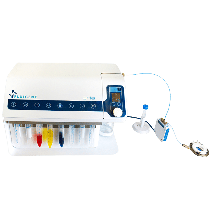
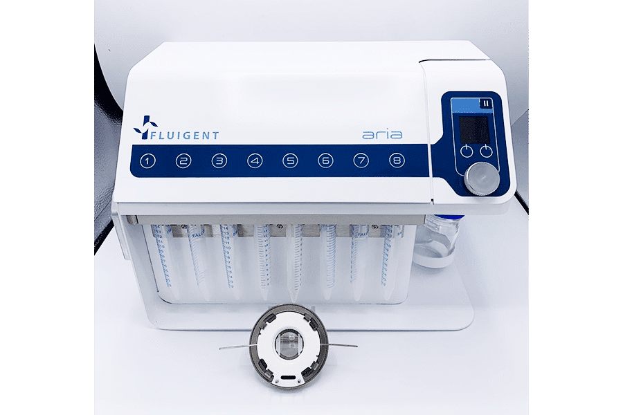
Fluigent has partnered with Bioptechs, the world leader in micro-environmental control chambers, to offer complete solutions for high-quality automated multiplexed imaging. The system combines a precision flow cell with a high-performance perfusion instrument. This flexible platform allows users to automate complex protocols with multiple fluid delivery for imaging.
Main benefits
 Flexible
- Time saving
Automation to gain productivity
- Compact
Easily moved between microscopes
Features of the platform
Preserve the sample
Temperature controlled flow cell: the flow cell can be cooled or heated from 0° – 58° C with less than 0.2° C variance and an integrated intelligent feedback loop for external temperature variables.
Controlled perfusion: avoids abrupt manual injection that can damage both the sample, the reagent, and the microscopy workstation.
Enclosed system: contamination free.
Reduce variability
Protocol automation drastically reduces variability to as little as 0.5% between experiments, compared to 5.1% intra-operator variability and 8.1% inter-operator variability using a pipette.
Synchronize with microscope
Aria and the FCS Chamber System are equipped with TTL ports to either send or receive TTL signals. TTL communication with a microscope enables users to synchronize perfusion and imaging.
Cost-effective
This automated multiplexed imaging platform is a cost-effective alternative to an all-in-one system. In addition, because the platform is flexible and transportable, it can be easily moved between microscopes to perform various sets of experiments, and is highly adaptable to different protocols and investigations.
What is multiplexed imaging?
Multiplexed imaging refers to a technique used in biological imaging to simultaneously visualize multiple targets or biomarkers within a sample. It allows researchers to obtain spatial and molecular information from a single sample, providing a more comprehensive understanding of complex biological systems. In traditional imaging techniques, such as immunofluorescence or immunohistochemistry, only a limited number of targets can be visualized at a time using specific fluorescent labels or antibodies. Multiplexed imaging expands the capability by enabling the detection of numerous targets in a single experiment.
There are various approaches to achieve multiplexed imaging. One common method involves using different fluorophores or fluorescent dyes that emit distinct wavelengths of light. By selecting fluorophores with non-overlapping emission spectra, multiple targets can be labeled and detected simultaneously. This technique is often referred to as multiplex fluorescence imaging.
Multiplexed imaging has significantly advanced our understanding of complex biological processes, such as cell signaling, tissue microenvironments, and disease pathology. By enabling the simultaneous detection of multiple targets within a sample, researchers can gain deeper insights into the spatial relationships and interactions between different biomolecules, cells, or tissues, leading to more comprehensive and detailed biological analyses.
Description of the multiple fluid delivery platform
The automated multiplexed imaging platform is the perfect compromise between manual pipetting and an all-in-one system dedicated to one specific application, integrating a microscope, a specific chip type, and a given set of solutions. Any protocol with multiple solution delivery can be automated, saving the scientist time and reducing variability between experiments compared to manual procedures.
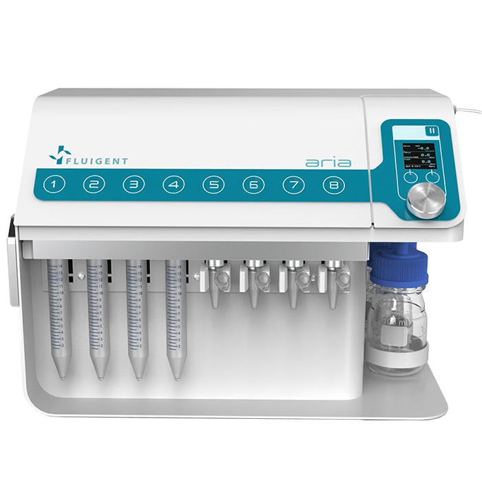
The platform, which integrates our ARIA automated sequential injection system, is straightforward and ready to use. No specific training is required, and no time is lost troubleshooting incidents or manually priming solutions before the experiment as with custom-built systems.
Improve your lab work with enhanced flexibility
The multiple fluid delivery platform is designed to offer maximum flexibility in terms of:
- Applications: the instrument and its intuitive software automates any sequence of functions: perfusion of up to 10 solutions, incubation, synchronization with the microscope via TTL signaling, flush tubing.
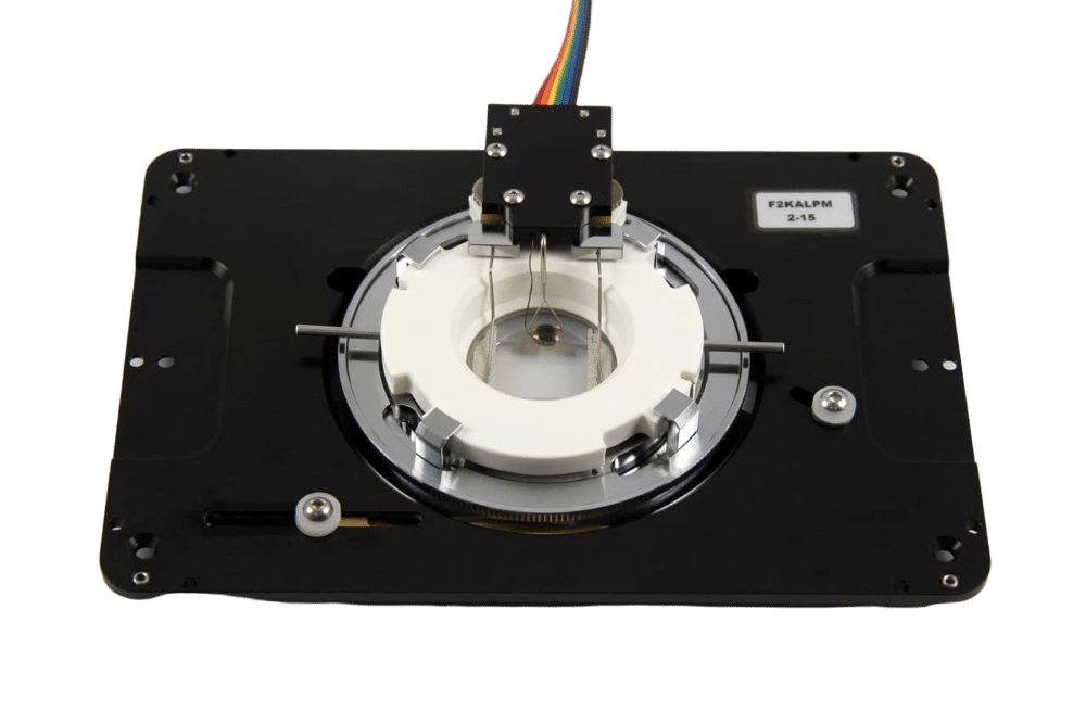
- Chamber design: the flow cell comes with a wide range of gaskets that fit biological samples from 2D cells to thick tissue samples. It is available in cooled, no-heat, and temperature-controlled versions, with an intelligent feedback system to maintain perfect temperature control for long-term investigations, and features leakproof flow.
- Microscopy: Our automated multiplexed imaging platform is adaptable to any upright or inverted microscope.
- Reagents: any type of antibody, RNA or DNA sequence can be used.

Productivity
Automation offers high productivity gains compared to manual pipetting.
The 20-step immunoassay protocol, which takes 2 days with manual pipetting, is shortened to several hours, increasing overall productivity from 2 samples a week to 1 sample a day. Automation also significantly reduces operational variability and pipetting errors.
Watch the webinar on the benefits of automation and examples of applications.
Applications of the multiple fluid delivery platform
This kit is perfect for any researcher who is looking to automate the labeling process for living cells, fixed tissues, and more.
Applications to which this system can be directly applied include but are not limited to:
- Shear stress experiments
- Extended imaging of live 3D cell culture
- Omics applications, including but not limited to proteomics and spatial-omics
- Ratiometric imaging such as calcium rationing
- Intracellular trafficking
- Any manner of fluorescence in situ labeling, including but not limited to FISH (fluorescence in situ hybridization), seq-FISH, MER-FISH, HCR-FISH
- Toxicity testing
- Drug response studies
- High-throughput expression screening
WEBINAR REPLAY – Automated IBEX multiplex immunohistochemistry with Aria
Join Dr. Colin Chu from the UCL Institute of Ophthalmology (UK) in this seminar, as he presents a powerful technique developed with the Germain Research Group at the US NIH: Iterative Bleaching Extends Multiplexity.
Using Fluigent’s Aria automated perfusion system, the team successfully labeled up to 40 proteins in a single tissue section—pushing the boundaries of multiplex imaging with microfluidics.
Webinar hosted by:
Amar Tamra, PhD, Application Engineer, Fluigent
Dr. Colin Chu, University College London
WEBINAR REPLAY – Automated fluid delivery for cell and tissue imaging
Want to gain time, precision and reproducibility with your immunofluorescence assay, or any other assay requiring injection of multiple solutions on your sample? Watch our webinar to learn how to interface a flow chamber (or a microfluidic chip) to our automated fluid delivery device ARIA. This will allow you to deliver up to 10 different solutions in a sequential and autonomous manner. In addition, the automated multiplexed imaging platform can be synchronized with any microscope to launch an image acquisition cycle and resume the perfusion protocol once the imaging cycle has been completed.
This all-in-one workflow facilitates alternation between cycles of injection/incubation time with reagents and image acquisition, a feature particularly well-adapted to complex cell and tissue imaging. Here we will present some example applications such as multiplexed tissue imaging, DNA-PAINT and seqFISH as well as cell capture and staining.
Webinar contents:
Reasons for & advantages of using automated sequential fluid delivery
Demo video
Example applications:
- Automated multiplexed tissue imaging
- Sequential fluorescence in situ hybridization (SeqFISH)
- Capture and characterization of circulating tumor cells
Q&A session
Webinar hosted by:
Mélanie Chabaud (Fluigent – Application Engineer)
William César (Fluigent – R&D project manager)
Noé Viovy (Fluigent – R&D software developer)
WEBINAR REPLAY – How to turn your fluorescence microscope into a spatial omics platform
Current approaches in genomics, transcriptomics, and proteomics yield quantitative abundance analysis of biomolecules on an almost routine basis, with a critical impact in the life sciences.
However, coupling this high content to spatial information in a single-cell and tissue context is still a challenge, and this is where our efforts are presently focusing.
In this webinar, I will share my facility’s experience in building spatial omics platforms.
First, I will provide an overview and comparison of microscopy-based methods for spatial omics. Then, I will present details and resources on how to build such a platform.
Finally, I will show the existing tools available for image and data analysis associated with these methods.
It is my hope that our experience can serve others in the road ahead to help decide which technique is best for their applications and to assist in their implementation.
Webinar hosted by:
Alvaro Crevenna: Head of Microscopy, EMBL Rome
Dale Clark: Chief Operating Officer, Bioptechs Inc.
Specifications
Aria software
Aria comes with an intuitive software that enables to design protocols in few clicks.
Incubation time, flow rate, volume dispensed, are all parameters that can be easily set by the operator for each step of his protocol. Protocols are recorded and can be shared among users. Flow rate and pressure are recorded for each experiment
Flow cell
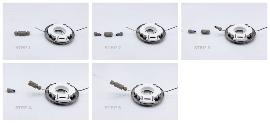
How to mount the flow cell?
How to connect Aria to the FCS2?
Connect the output of the 2 switch valve to the flow cell using a P702 union
Watch this step-by-step tutorial series to master Aria — from setup to full software control.
Episode 1
Episode 2
Episode 3
Episode 4
Episode 5
Episode 6
Episode 7