In this application note we are investigating the usability of the commercially available surfactant dSurf for an exemplary digital PCR-assay.
Thanks to droplet microfluidic, Michael Ryckelynck and his team are able to isolate single DNA molecules and analyze the enzymes and proteins resulting from their expression.
We discuss some applications where microfluidics achieves results that would be very challenging to obtain when using conventional methods.
Microfluidic methods can be used to improve vaccine research and development. Microfluidic techniques are already used to develop adjuvants, perform virus identification/diagnostics, or drug micro and nanoencapsulation.
Fluigent has developed a new system to overcome the limitations of common designs used in droplet generation chips.
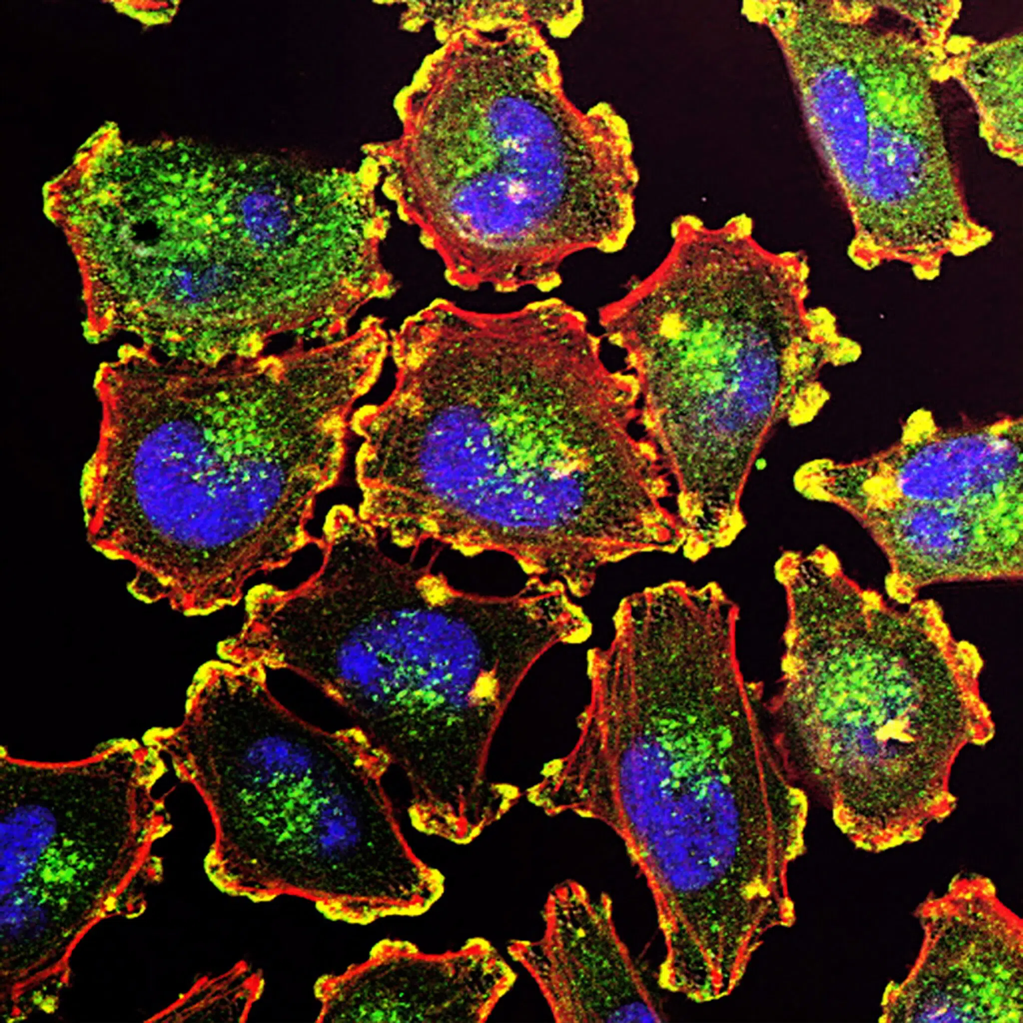
Micropipette aspiration is a powerful non-invasive technique to evaluate how biomechanical properties of single cells or tissue govern cell shape, cell response to mechanic stimuli, transition from nontumorigenic to tumorigenic state or morphogenesis
The choice of the right method of fluid management can often determinate the success of a project, as well as the overall time and cost of manufacturing. Here is what you need to consider and be aware of when integrating fluidic control into your system.
Complete and in-depth overview on droplet microfluidics.
Many national and internationally funded projects bring together science and innovation. These projects cover a large number of subjects. Fluigent takes part in several European and French funded programs to provide expertise and resources in microfluidics and related applications.
Droplet microfluidics is a powerful tool which consists of generating and manipulating micron-sized monodispersed droplets.
In this application note, we present droplet generation data obtained using decane in Water, a system that demonstrates the expected behavior of most hydrocarbons in Water. We demonstrate the ability of Fluigent equipment coupled with Raydrop microfluidic devices to generate high-quality emulsions with controlled droplet sizes and with high throughput.
In this application note, we present droplet generation data obtained using one of the most widely used water in oil emulsion systems – water in decane. We demonstrate the ability of Fluigent equipment coupled with RayDrop microfluidic devices to generate high-quality emulsions with controlled droplet sizes and with high throughput.
Achieve reproducible, high-quality water in fluorocarbon oil emulsions with cutting-edge microfluidic devices. Ideal for Drop-Seq and RNA-Seq studies.
The Drop-Seq protocol, is a high throughput method that enables the sequencing of the mRNA from a large number of cells. With this method it is possible to create a gene expression map of the cell, or even distinguish cell populations within a tissue!
A microscope designed for microfluidics
Microfluidic Droplet Pack
P-OEM
OEM Fluigent PX
Chip for electrochemical gradients to 3D cell cultures