Cartilage on chip using Fluigent MFCS pressure controller

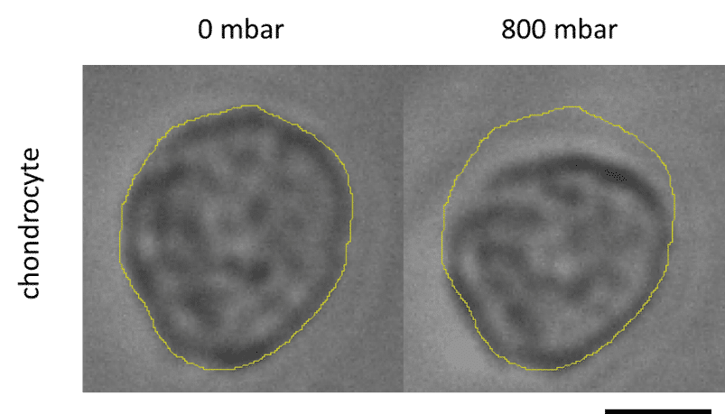
Elucidating how chondrocytes react to external stimuli (mechanical or chemical) is important to understand processes triggering cartilage diseases like osteoarthritis. To this end, various approaches, have been explored so far, while they all suffer from specific limitations.

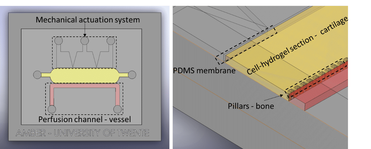
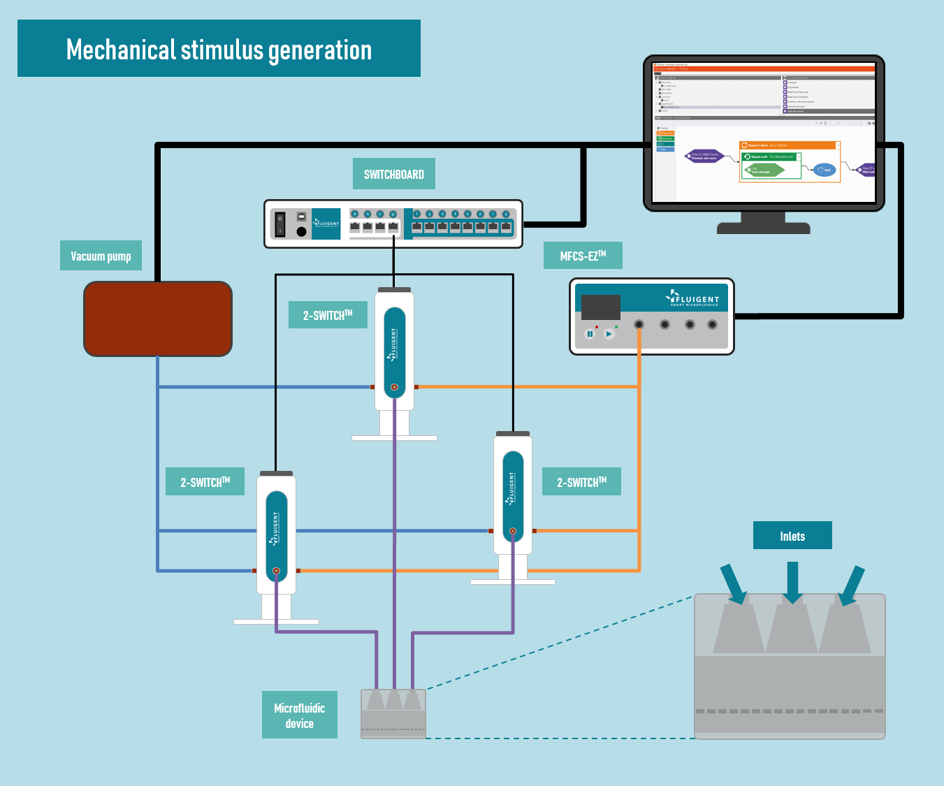
In this application note, we report on the use of Fluigent products to create complex mechanical stimulation patterns on 3D cell culture in a microfluidic platform, or so-called organ-on-a-chip device, with a specific focus on creating a cartilage-on-a-chip model.


This application note was made possible thanks to Severine Le Gac and her team at Applied Microfluidics for BioEngineering Research (AMBER). For more information visit the team website.

INTRODUCTION

schema-compressive-stress-cell

Articular cartilage covers the bone friction surfaces in joints (Fig. on the left). The principal function of the cartilage is to provide a smooth, lubricated surface for articulation and to facilitate the transmission of loads between the bones.

Current method for study how chondrocytes react to external stimuli include 2D or 3D culture. Both have limitations including non-physiological phenotype, failing to provide a dynamic cell microenvironment as found in vivo, or more importantly specific mechanical stimulation.

To create more advanced culture platforms and address some of the aforementioned limitations, microfluidics has been introduced. It notably allows implementing mechanical stimulation on 3D cell culture and studying in situ the cell response to these stimulations, as well as creating dynamic culture conditions.


Partial materials and methods

Compressive stress

Microfluidic platform

The plateforme was fabricated using soft-lithography in PDMS, and assembled to a PDMS-coated glass slide. This platform allows replicating the following main cartilage parts and functions with a culture chamber, a nutrient supply and a mechanical stress

Mechanical stimulus generation

A MFCS-EZ pressure controller coupled to three 2-switch valves has been used to generate the mechanical stimulation in the cartilage-on-a-chip platform (Compression and/or shear stress). Fluigent automation software was used to automate the process and produce complex stimulation patterns, by changing the type of stimulus (positive or negative pressure) and its amplitude applied to the three actuation chambers (Figure 3).

A camera (ORCA-flash 4.0 LT, Hamamatsu Photonics) was used to monitor and analyse the PDMS membrane deformation upon application of various pressure patterns (negative or positive pressure, the latter ranging from 0 mbar to 1500 mbar).

mimic compression
Cartilage-OAC

Partial results


Cell culture experiments

Chondrocytes were cultured in the microfluidic platform for 6 days under static (fig. below) or dynamic conditions. Dynamic conditions were applied in the form of 1.5 h of stimulation of the cell-laden hydrogel per day, using uniform compression (application of 800 mbar on all three actuation chambers with a 1-Hz frequency).

Furthermore, the cell deformation across the chamber was characterized (Fig.); it amounted to 13% close to the membrane, and decreased across the cell culture chamber.

Chondrocyte deformation
Figure: Chondrocyte deformation: a) no pressure; b) 800 mbar of compression. (Scale: 7µm)


Conclusion

In the past years, transitioning from traditional 2D culture to 3D culture was a big improvement for in vitro systems. The example illustrated in this application note is the use of microfluidic to reproduce the mechanical forces exerted on the articular cartilage. Fluigent products were decisive to reproduce such stimulation. Their precision and the ability to fully automate different fluidic component at once were determinant to reproduce the amplitude and frequency of stimulation over long term experiment.


Related products

For more information or a technical discussion

Contact us