No tools needed
Live Cell Imaging Chamber
FCS2 Chamber
The Focht Chamber System 2 (FCS2®) is a closed system live-cell micro-observation chamber that operates using several patented technologies that provide various exclusive advantages over alternative chambers. In addition to its uniform temperature control and user-definable perfusion capability, it is fully compatible with all microscopy modes. The chamber adapts to protocol instead of having to modify your procedure to fit the chamber. to fit the chamber. the chamber.

Main benefits
 Easy
Easy Flexible
Fits with all microscopes
 Control
Control all parameters
FCS2 Live Cellular Imaging Features
Complete control
The live cell imaging chip FCS2 provides precise control over the volume within the optical cavity, the separation between optical surfaces, and of the shape or profile of the flow channel. A highly efficient, fast, and uniform temperature control, both above and below ambient conditions.
Dedicated to cell imaging
The FCS2 is purposely built for live cell imaging applications. The FCS2 is designed to accommodate adherent, tissue samples, or suspended specimens to deliver high quality results in capturing dynamic cellular processes.
Adaptable
Fluigent’s live cell imaging culture chamber adapts to all microscope brands, and is fully compatible with all microscopy modes.
Related applications
Description
The FCS2 is a flow cell or flow chamber that is designed specifically for the demands of today’s live-cell imaging requirements. This live cell imaging flow chamber has limitless flow characteristics because its flow geometry can easily be customized by the user. The most important feature of the FCS2, other than unequalled temperature uniformity, is that the flow of media is constrained to a precise location over the cells. The FCS2 provides unmatched, uniform temperature control, in a user configurable, perfusable, optical imaging cavity.
FCS2 Chamber
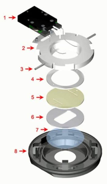
The FCS2 flow chamber components:
- Electrical Enclosure
Can also be detached to sterilize the perfusion tubes
Temperature sensor
Heater contacts - Upper Half (Top)
Contains the perfusion tubes - Perfusion Tubes (14 gauge) (CO2 Perfusion Drawing)
- Upper Gasket
- Microaqueduct Slide
An optical surface which integrates perfusion and temperature control
High-volume laminar flow
Koehler Illumination
Electronically conductive coating for temperature control - Singular lower gasket
This gasket can have any internal geometry you desire
Standard thicknesses from .1mm to 1mm
Allows you to define the volume and flow characteristics of the chamber - 40mm coverslip
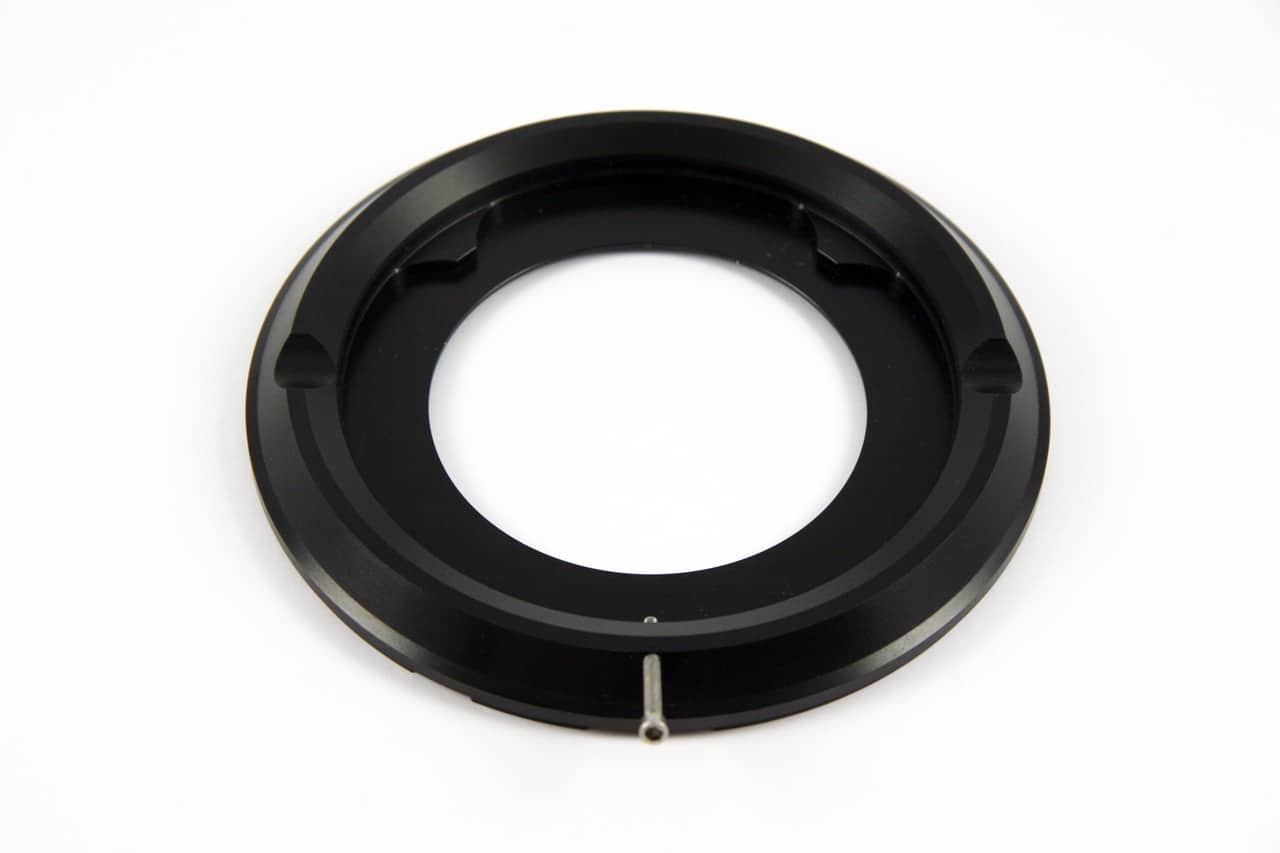
Surface where your cells are grown - Self locking base
Designed to assure parallel uniform closure, eliminate leaks, & broken coverslips
Temperature controlled
Dovetail mounted to scope for stability
No tools for assembly
Items 5,6 & 7 of the live cell imaging culture chamber are the optical cavity, the gasket (#6) can be changed to anyone to make any flow geometry, or media volume you want. The coverslip comes in a standard 0.17mm, #1.5 thickness but is also available in 0.5mm, #5 coverslip.
Cells are grown on a 40mm glass coverslip. This coverslip is then incorporated into a perfusable fluid optical cavity that is compatible with all modes of microscopy, and its geometry can be easily defined by the user. This optical cavity is secured into a fixture on the stage of the microscope where it can be perfused with media or remain static.
Media that comes into one of the ports on the side of the live cell imaging chamber, emerges in a fluid optical path where the media is precisely directed over the cells. The media is collected within the optical cavity and directed out of the chamber on the other side.
Customized perfusion holes
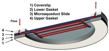
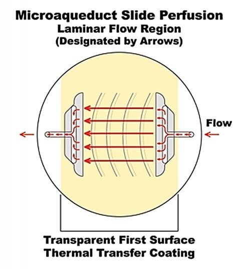
Microaqueduct Perfusion: uniform laminar flow perfusion in the whole live cellular imaging chamber
A fluid pathway is formed by separating the Microaqueduct Slide (3) from the coverslip (1) containing cells with a single silicone gasket (2). This gasket (2) can be any thickness from 50 micron to 1mm and any lateral geometry you choose or create. This arrangement allows the user to define the flow characteristics.
Therefore, you are not limited by the geometry of the optical cavity. Instead, you select or create it! Fluid access to this flow channel is made through two 14-gauge needle stock tubes protruding from the sides of the chamber top.
These tubes provide fluid connection to two perfusion holes in the Microaqueduct Slide that interface two “T” shaped grooves cut into the inner surface of the Microaqueduct Slide. The “T” groove allows the media to seek the path of least resistance and become nearly laminar before flowing across the cells.
This technique eliminates the need for the metal perfusion ring and additional gaskets, which are the limiting factors required by most conventional chambers.
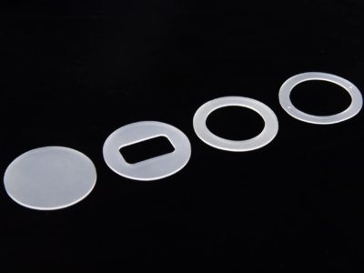
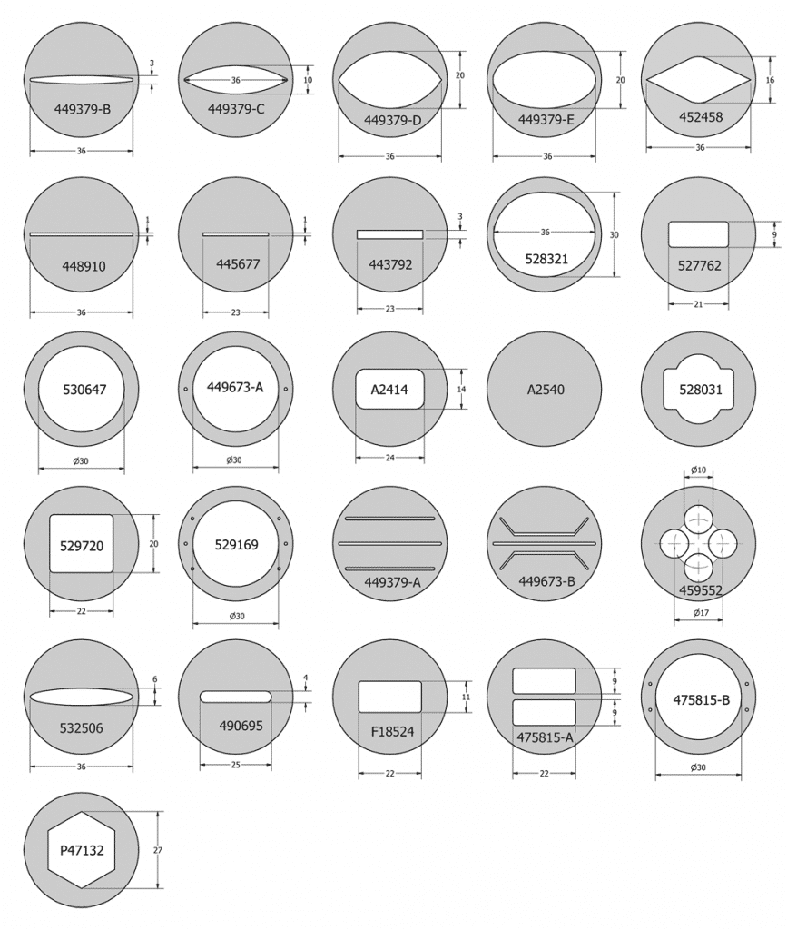
Singular lower gasket: Find the gasket that meets your experiment requirements
By simply changing this one gasket you can change the volume of the live cellular imaging chamber. This gasket can have any internal geometry you desire and can be any thickness from 0.1mm to 1mm. The geometry of the gasket allows to constrain the flow of media to a precise location over the cells. Blank, rectangle and round gaskets are standard geometries, but the gasket can be customized to fit your application.
Temperature controller
The FCS2 was designed to maintain precise thermal control. The flow cell can be cooled down or heated up from 0 – 58 C with less than 0.2 C variance and integrated intelligent feedback loop for external temperature variables. Temperature variations can be set and synchronized with imaging or perfusion via TTL signaling.
The surface of the Microaqueduct Slide, opposite the specimen side, is coated with an electrically conductive transparent thin film of Indium-Tin Oxide (ITO) and two electrical contacts (busbars). When the FCS2 is completely assembled, two electrical contacts, which are contained in the electrical enclosure, rest on the busbars. A temperature controller is used to pass a regulated current flow through the ITO Coating. This causes the surface of the slide to heat. The heat is transferred through the perfusable media to the cell surface on the coverslip there by providing first surface thermal control. The self-locking base of the live cell imaging system is also temperature regulated to provide peripheral heat as well.
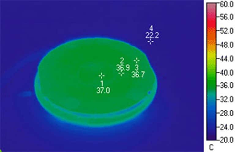
This thermographic image demonstrates the uniform temperature distribution of an FCS2 live cellular imaging. Notice that the coverslip temperature is so uniform that its location, in infrared, is indistinguishable from the base of the chamber.
This demonstrates the effectiveness of the ITO heated Microaqueduct Slide. It is capable of re-equilibrating cell temperature within seconds of perfusion and eliminates the typical thermal gradient that occurs with peripheral heating.
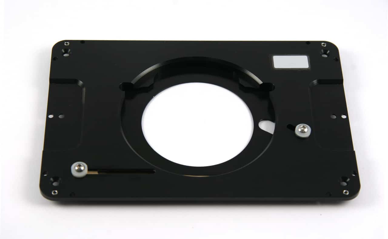
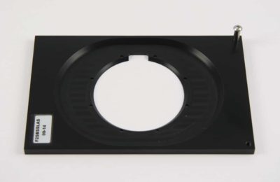
Stage adaptors for FCS2
The Bioptechs live cellular imaging FCS2 Stage Adapter interfaces the microscope stage with the FCS2 chamber.
If you do not see a stage adapter that will fit your stage, reach out. Bioptechs is always adding to its catalog of stage geometries and specifications.
Standard stage adapters available for FCS2 live cell imaging solution:
Live Cell Imaging Chamber FCS2 Specifications
FCS2 Chamber loading