Capture and Labeling of Cancer Cells Using Aria

The invasiveness of cancer cells describes the metastasizing capability of a primary tumor. The straightforward detection and quantification of cancer cell invasion are important to predict the survival rate of a cancer patient and to test how anti-cancer compounds influence cancer progression.

This application note describes the use of the new Fluigent Aria – a software-assisted instrument capable of delivering up to 10 different solutions – for the automation of a complete protocol of capture and labelling of MDA-MB-231 breast cancer cells including surface treatment, injection of antibodies-coated beads, cell suspension, and immunostaining steps. The Aria creates an automated cancer cell analysis, thus saving time and reagents while considerably reducing margins of error.

We have demonstrated the use of Aria and its related software for the automated delivery of different liquids for the capture and labeling of breast cancer cells using a complex microfluidic setup. We also demonstrate how Aria can adapt to specific protocols via its software features, allowing for the optimization of protocols.

Context

Cancer is the second leading cause of death worldwide. Greater than 90% of deaths in cancer patients are attributed to metastasis. Cancer is considered a localized disease in its early stage. However, it has often become systemic by the time a patient becomes symptomatic and the disease is detected by currently available imaging modalities such as traditional radiography (X-ray), magnetic resonance imaging (MRI), computed tomography (CT), positron emission tomography (PET), or ultrasound. There is growing evidence that cancer cells are shed from the primary tumor into the circulation prior to the presentation of clinical symptoms. These circulating tumor cells (CTCs) may finally colonize at distant sites and form metastases.

Since CTCs are mainly characterized by their morphology and immunostaining pattern, their heterogeneity is a major obstacle for CTC detection. The CTCs derived from different types of tissues significantly distinguish from each other with different size, shape, and immunophenotyping profiles. However, there is broad morphological and immunophenotypical variation within CTCs derived from the same tissue of origin.

Cell preparation and antibody staining for CTC cell detection is time consuming and labour intensive. The process can last for several hours and often requires exposure to multiple fluids.  For example, typical staining protocols usually require more than 5 consecutive solutions.

To carry out reproducible investigations, accurate injection volumes and controlled flow rates (between 1 and 1000 µL/min) are necessary for cell labeling, cell manipulation, drug screening, or perfusion. Smooth fluid control is also recommended since severe shear stresses caused by unregulated flow rates might change the functional characteristics of cells and damage samples. Aria was designed to overcome these issues and automate fluid supply procedures to obtain an automated cancer cell analysis.

Automated Cancer Cell Analysis: Challenge

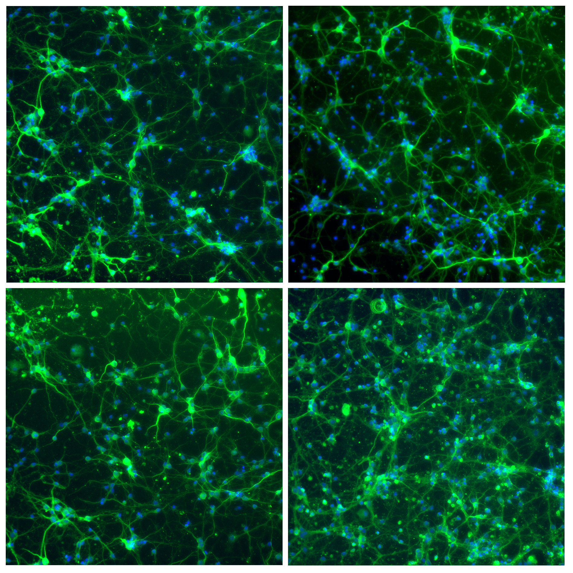
We describe the automation of a rare cell capture and detection method including surface treatment, beads and cells solution injections, and immunostaining using precise volume control using Aria and its related automation software. We use the EPHESIA microfluidic chip from Institut Curie and MDA-MB-231 breast cancer cell line to demonstrate system function and utility.

AUTOMATED CANCER CELL ANALYSIS setup

Conclusion

Aria and its related software were used for the automated delivery of different liquids for the capture and labelling of breast cancer cells using a complex microfluidic setup. This protocol included 10 different liquid injections, surface treatment, beads injection, cells injection, capturing,  and labelling in a sequential and automated manner. Thus, we have demonstrated the use of Aria for optimal automated cancer cell analysis.

Aria can also adapt to specific protocols by making use of its specific software features, allowing for the optimization of complex protocols.

Aria provides key features for performing this application as it brings to the user:

  • Reduced manipulation time and timed exposure to antibodies, fluorophores, and DNA probes with automated and timed protocols
  • Reduced handling for minimal contamination< or changes to cell conditions
  • Higher reproducibility compared to manual methods, resulting in more reliable results

References

  1. Autebert, J. et al. 2015 High purity microfluidic sorting and analysis of circulating tumor cells: towards routine mutation detection. Lab Chip 15, 2090–2101 (2015).
  2. Kim, M. Y. et al. Tumor Self-Seeding by Circulating Cancer Cells. Cell 139, 1315–1326 (2009).
  3. Cabel, L. et al. Clinical potential of circulating tumour DNA in patients receiving anticancer immunotherapy. Nat. Rev. Clin. Oncol. 15, 639–650 (2018).
  4. Bidard, F. C. et al. Clinical validity of circulating tumour cells in patients with me- tastatic breast cancer: A pooled analysis of individual patient data. Lancet Oncol. 15, 406–414 (2014).
  5. Saias, L., Autebert, J., Malaquin, L. & Viovy, J.-L. 2011 Design, modeling and characte- rization of microfluidic architectures for high flow rate, small footprint microfluidic systems. Lab Chip 11, 822–32 (2011).
  6. Bernacka-wojcik, I. 2014 Design and development of a microfluidic platform for use with colorimetric gold nanoprobe assays. (Universidade Nova de Lisboa, 2014)

For more information or a technical discussion

Contact us